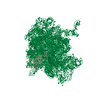
Movie
Movie Controller


+ Open data

- Basic information
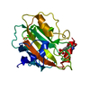
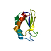
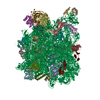
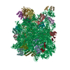
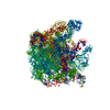
| Entry | Database: PDB / ID: 4ce4 | ||||||||||||
|---|---|---|---|---|---|---|---|---|---|---|---|---|---|
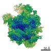
| Title | 39S large subunit of the porcine mitochondrial ribosome | ||||||||||||
 Components Components |
| ||||||||||||
 Keywords Keywords | RIBOSOME / MAMMALIAN MITOCHONDRIAL RIBOSOME / 39S LARGE RIBOSOMAL SUBUNIT / TRANSLATION / RIBOSOMAL PROTEINS / RRNA / POLYPEPTIDE EXIT SITE / MEMBRANE ASSOCIATION | ||||||||||||
| Function / homology |  Function and homology information Function and homology informationMitochondrial translation elongation / Mitochondrial ribosome-associated quality control / rRNA import into mitochondrion / mitochondrial translational termination / ribonuclease III activity / mitochondrial translational elongation / translation release factor activity, codon nonspecific / Mitochondrial translation termination / Mitochondrial protein degradation / peptidyl-tRNA hydrolase ...Mitochondrial translation elongation / Mitochondrial ribosome-associated quality control / rRNA import into mitochondrion / mitochondrial translational termination / ribonuclease III activity / mitochondrial translational elongation / translation release factor activity, codon nonspecific / Mitochondrial translation termination / Mitochondrial protein degradation / peptidyl-tRNA hydrolase / mitochondrial large ribosomal subunit / peptidyl-tRNA hydrolase activity / mitochondrial translation / organelle membrane / RNA processing / double-stranded RNA binding / large ribosomal subunit / 5S rRNA binding / mitochondrial inner membrane / nuclear body / rRNA binding / structural constituent of ribosome / ribosome / translation / ribonucleoprotein complex / protein domain specific binding / nucleotide binding / mitochondrion / RNA binding / nucleus / plasma membrane / cytosol / cytoplasm Similarity search - Function | ||||||||||||
| Biological species |  | ||||||||||||
| Method | ELECTRON MICROSCOPY / single particle reconstruction / cryo EM / Resolution: 4.9 Å | ||||||||||||
 Authors Authors | Greber, B.J. / Boehringer, D. / Leitner, A. / Bieri, P. / Voigts-Hoffmann, F. / Erzberger, J.P. / Leibundgut, M. / Aebersold, R. / Ban, N. | ||||||||||||
 Citation Citation | Journal: Nature / Year: 2014 Title: Architecture of the large subunit of the mammalian mitochondrial ribosome. Authors: Basil J Greber / Daniel Boehringer / Alexander Leitner / Philipp Bieri / Felix Voigts-Hoffmann / Jan P Erzberger / Marc Leibundgut / Ruedi Aebersold / Nenad Ban /  Abstract: Mitochondrial ribosomes synthesize a number of highly hydrophobic proteins encoded on the genome of mitochondria, the organelles in eukaryotic cells that are responsible for energy conversion by ...Mitochondrial ribosomes synthesize a number of highly hydrophobic proteins encoded on the genome of mitochondria, the organelles in eukaryotic cells that are responsible for energy conversion by oxidative phosphorylation. The ribosomes in mammalian mitochondria have undergone massive structural changes throughout their evolution, including ribosomal RNA shortening and acquisition of mitochondria-specific ribosomal proteins. Here we present the three-dimensional structure of the 39S large subunit of the porcine mitochondrial ribosome determined by cryo-electron microscopy at 4.9 Å resolution. The structure, combined with data from chemical crosslinking and mass spectrometry experiments, reveals the unique features of the 39S subunit at near-atomic resolution and provides detailed insight into the architecture of the polypeptide exit site. This region of the mitochondrial ribosome has been considerably remodelled compared to its bacterial counterpart, providing a specialized platform for the synthesis and membrane insertion of the highly hydrophobic protein components of the respiratory chain. | ||||||||||||
| History |
| ||||||||||||
| Remark 700 | SHEET DETERMINATION METHOD: DSSP THE SHEETS PRESENTED AS "EA" IN EACH CHAIN ON SHEET RECORDS BELOW ... SHEET DETERMINATION METHOD: DSSP THE SHEETS PRESENTED AS "EA" IN EACH CHAIN ON SHEET RECORDS BELOW IS ACTUALLY AN 6-STRANDED BARREL THIS IS REPRESENTED BY A 7-STRANDED SHEET IN WHICH THE FIRST AND LAST STRANDS ARE IDENTICAL. |
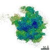
- Structure visualization
| Movie |
Movie viewer |
|---|---|
| Structure viewer | Molecule: MolmilJmol/JSmol |
- Downloads & links
-Download
| PDBx/mmCIF format | 4ce4.cif.gz | 1.4 MB | Display | PDBx/mmCIF format |
|---|---|---|---|---|
| PDB format | pdb4ce4.ent.gz | 1.1 MB | Display | PDB format |
| PDBx/mmJSON format | 4ce4.json.gz | Tree view | PDBx/mmJSON format | |
| Others |  Other downloads Other downloads |
-Validation report
| Arichive directory | https://data.pdbj.org/pub/pdb/validation_reports/ce/4ce4ftp://data.pdbj.org/pub/pdb/validation_reports/ce/4ce4 | HTTPS FTP |
|---|
-Related structure data
| Related structure data | 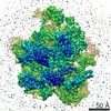 2490MC M: map data used to model this data C: citing same article ( |
|---|---|
| Similar structure data |

-Links
 PDBj
PDBj


- Assembly
| Deposited unit | 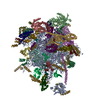
|
|---|---|
| 1 |
|
-Components
+Protein , 35 types, 36 molecules 012356789DEFINOPQRSTUVWXYbchil...
-RNA chain , 2 types, 2 molecules AB
| #10: RNA chain | Mass: 501424.250 Da / Num. of mol.: 1 / Source method: isolated from a natural source / Source: (natural) |
|---|---|
| #11: RNA chain | Mass: 5641.792 Da / Num. of mol.: 1 / Source method: isolated from a natural source / Source: (natural) |
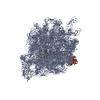
-Non-polymers , 1 types, 2 molecules 
| #38: Chemical |  Mass: 65.409 Da / Num. of mol.: 2 / Source method: obtained synthetically / Formula: Zn Mass: 65.409 Da / Num. of mol.: 2 / Source method: obtained synthetically / Formula: Zn |
|---|
-Details
| Sequence details | CHAINS I W X 7: ACCESSION CODE PROVIDED FOR MRNA SEQUENCE, PROTEIN SEQUENCE IS TRANSLATED MRNA. ...CHAINS I W X 7: ACCESSION CODE PROVIDED FOR MRNA SEQUENCE, PROTEIN SEQUENCE IS TRANSLATED |
|---|
-Experimental details
-Experiment
| Experiment | Method: ELECTRON MICROSCOPY |
|---|---|
| EM experiment | Aggregation state: PARTICLE / 3D reconstruction method: single particle reconstruction |
- Sample preparation
| Component | Name: 39S MITOCHONDRIAL RIBOSOMAL SUBUNIT / Type: RIBOSOME |
|---|---|
| Buffer solution | Name: 20 MM HEPES-KOH PH 7.9, 100 MM KCL, 1 MM DTT, 20 MM MGCL2 pH: 7.9 Details: 20 MM HEPES-KOH PH 7.9, 100 MM KCL, 1 MM DTT, 20 MM MGCL2 |
| Specimen | Conc.: 0.07 mg/ml / Embedding applied: NO / Shadowing applied: NO / Staining applied: NO / Vitrification applied: YES |
| Specimen support | Details: HOLEY CARBON |
| Vitrification | Instrument: HOMEMADE PLUNGER / Cryogen name: ETHANE Details: MANUAL BLOTTING FOLLOWED BY MANUAL PLUNGING INTO LIQUID ETHANE |
- Electron microscopy imaging
| Experimental equipment |  Model: Titan Krios / Image courtesy: FEI Company |
|---|---|
| Microscopy | Model: FEI TITAN KRIOS Details: IMAGES COLLECTED ON A FEI TITAN KRIOS BETWEEN NOVEMBER 2012 AND JUNE 2013. CALIBRATED MAGNIFICATION 141510 FOR FALCON II DATA AND 99290 FOR FALCON I DATA. |
| Electron gun | Electron source:  FIELD EMISSION GUN / Accelerating voltage: 300 kV / Illumination mode: FLOOD BEAM FIELD EMISSION GUN / Accelerating voltage: 300 kV / Illumination mode: FLOOD BEAM |
| Electron lens | Mode: BRIGHT FIELD / Nominal magnification: 59000 X / Nominal defocus max: 3600 nm / Nominal defocus min: 1200 nm / Cs: 2.7 mm |
| Specimen holder | Temperature: 79 K / Tilt angle max: 0 ° |
| Image recording | Electron dose: 20 e/Å2 / Film or detector model: FEI FALCON II (4k x 4k) |
| Radiation wavelength | Relative weight: 1 |
- Processing
| EM software |
| ||||||||||||||||||||||||||||||||||||||||||||||||||||||||||||||||||||||
|---|---|---|---|---|---|---|---|---|---|---|---|---|---|---|---|---|---|---|---|---|---|---|---|---|---|---|---|---|---|---|---|---|---|---|---|---|---|---|---|---|---|---|---|---|---|---|---|---|---|---|---|---|---|---|---|---|---|---|---|---|---|---|---|---|---|---|---|---|---|---|---|
| CTF correction | Details: PER DETECTOR FRAME | ||||||||||||||||||||||||||||||||||||||||||||||||||||||||||||||||||||||
| Symmetry | Point symmetry: C1 (asymmetric) | ||||||||||||||||||||||||||||||||||||||||||||||||||||||||||||||||||||||
| 3D reconstruction | Method: ANGULAR RECONSTITUTION FOR INITIAL MODEL GENERATION FOLLOWED BY REFINEMENT BY PROJECTION MATCHING Resolution: 4.9 Å / Resolution method: FSC 0.143 CUT-OFF / Num. of particles: 232181 / Actual pixel size: 1.41 Å Details: RESOLUTION DETERMIED ACCORDING TO GOLD STANDARD FSC BY FSC EQUAL 0.143 CRITERION FROM TWO INDEPENDENTLY REFINED DATA HALF SETS Refinement type: HALF-MAPS REFINED INDEPENDENTLY / Symmetry type: POINT | ||||||||||||||||||||||||||||||||||||||||||||||||||||||||||||||||||||||
| Atomic model building | Protocol: OTHER / Space: REAL Details: REFINEMENT PROTOCOL--X-RAY STRUCTURES, HOMOLOGY MODELS | ||||||||||||||||||||||||||||||||||||||||||||||||||||||||||||||||||||||
| Atomic model building |
| ||||||||||||||||||||||||||||||||||||||||||||||||||||||||||||||||||||||
| Refinement | Highest resolution: 4.9 Å | ||||||||||||||||||||||||||||||||||||||||||||||||||||||||||||||||||||||
| Refinement step | Cycle: LAST / Highest resolution: 4.9 Å
|