 Movie
Movie Controller

[English] 日本語
 Yorodumi
- PDB-5ntn: Structural states of RORgt: X-ray elucidation of molecular mechan... -
+ Open data

- Basic information
| Entry | Database: PDB / ID: 5ntn | ||||||
|---|---|---|---|---|---|---|---|
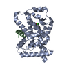
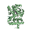
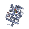
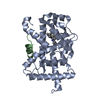
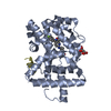
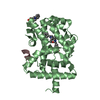
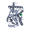
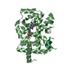
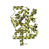
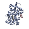
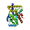
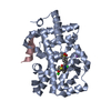
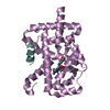
| Title | Structural states of RORgt: X-ray elucidation of molecular mechanisms and binding interactions for natural and synthetic compounds | ||||||
 Components Components |
| ||||||
 Keywords Keywords | SIGNALING PROTEIN / nuclear hormone receptor / ligand-binding domain / agonist | ||||||
| Function / homology |  Function and homology information Function and homology informationovarian follicle rupture / NR1H2 & NR1H3 regulate gene expression linked to gluconeogenesis / tolerance induction in gut-associated lymphoid tissue / T-helper 17 cell differentiation / regulation of steroid metabolic process / cellular response to sterol / nuclear glucocorticoid receptor binding / ligand-modulated transcription factor activity / Peyer's patch development / NR1H2 & NR1H3 regulate gene expression linked to lipogenesis ...ovarian follicle rupture / NR1H2 & NR1H3 regulate gene expression linked to gluconeogenesis / tolerance induction in gut-associated lymphoid tissue / T-helper 17 cell differentiation / regulation of steroid metabolic process / cellular response to sterol / nuclear glucocorticoid receptor binding / ligand-modulated transcription factor activity / Peyer's patch development / NR1H2 & NR1H3 regulate gene expression linked to lipogenesis / lipid storage / regulatory T cell differentiation / T-helper cell differentiation / positive regulation of circadian rhythm / RUNX3 Regulates Immune Response and Cell Migration / oxysterol binding / negative regulation of thymocyte apoptotic process / regulation of fat cell differentiation / Phosphorylated BMAL1:CLOCK (ARNTL:CLOCK) activates expression of core clock genes / histone deacetylase complex / regulation of glucose metabolic process / lymph node development / adipose tissue development / nuclear retinoid X receptor binding / xenobiotic metabolic process / RORA,B,C and NR1D1 (REV-ERBA) regulate gene expression / SUMOylation of transcription cofactors / Expression of BMAL (ARNTL), CLOCK, and NPAS2 / nuclear estrogen receptor binding / nuclear receptor binding / cellular response to estradiol stimulus / circadian regulation of gene expression / Heme signaling / circadian rhythm / Nuclear Receptor transcription pathway / DNA-binding transcription repressor activity, RNA polymerase II-specific / nuclear receptor activity / fibrillar center / histone deacetylase binding / sequence-specific double-stranded DNA binding / transcription corepressor activity / Interleukin-4 and Interleukin-13 signaling / Estrogen-dependent gene expression / DNA-binding transcription factor activity, RNA polymerase II-specific / transcription coactivator activity / nuclear speck / nuclear body / RNA polymerase II cis-regulatory region sequence-specific DNA binding / DNA-binding transcription factor activity / signaling receptor binding / regulation of transcription by RNA polymerase II / positive regulation of DNA-templated transcription / chromatin / nucleolus / negative regulation of transcription by RNA polymerase II / positive regulation of transcription by RNA polymerase II / zinc ion binding / nucleoplasm / nucleus / cytosol Similarity search - Function | ||||||
| Biological species |  Homo sapiens (human) Homo sapiens (human) | ||||||
| Method |  X-RAY DIFFRACTION / SYNCHROTRON / MOLECULAR REPLACEMENT / molecular replacement / Resolution: 1.9 Å X-RAY DIFFRACTION / SYNCHROTRON / MOLECULAR REPLACEMENT / molecular replacement / Resolution: 1.9 Å | ||||||
 Authors Authors | Kallen, J. | ||||||
 Citation Citation | Journal: ChemMedChem / Year: 2017 Title: Structural States of ROR gamma t: X-ray Elucidation of Molecular Mechanisms and Binding Interactions for Natural and Synthetic Compounds. Authors: Kallen, J. / Izaac, A. / Be, C. / Arista, L. / Orain, D. / Kaupmann, K. / Guntermann, C. / Hoegenauer, K. / Hintermann, S. | ||||||
| History |
|
- Structure visualization
| Structure viewer | Molecule: MolmilJmol/JSmol |
|---|
- Downloads & links
-Download
| PDBx/mmCIF format | 5ntn.cif.gz | 229.1 KB | Display | PDBx/mmCIF format |
|---|---|---|---|---|
| PDB format | pdb5ntn.ent.gz | 184.5 KB | Display | PDB format |
| PDBx/mmJSON format | 5ntn.json.gz | Tree view | PDBx/mmJSON format | |
| Others |  Other downloads Other downloads |
-Validation report
| Arichive directory | https://data.pdbj.org/pub/pdb/validation_reports/nt/5ntnftp://data.pdbj.org/pub/pdb/validation_reports/nt/5ntn | HTTPS FTP |
|---|
-Related structure data
| Related structure data | 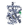 5ntiC 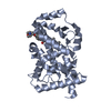 5ntkC 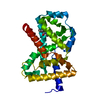 5ntpC 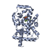 5ntqC 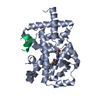 5ntwC  5nu1C 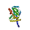 1s0xS S: Starting model for refinement C: citing same article ( |
|---|---|
| Similar structure data |


-Links
 PDBj
PDBj
- Assembly
| Deposited unit | 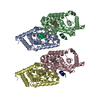
| ||||||||
|---|---|---|---|---|---|---|---|---|---|
| 1 | 
| ||||||||
| 2 | 
| ||||||||
| 3 | 
| ||||||||
| 4 | 
| ||||||||
| Unit cell |
|
-Components
| #1: Protein | Mass: 29739.316 Da / Num. of mol.: 4 / Fragment: C-terminal domain, ligand binding domain / Mutation: C455S Source method: isolated from a genetically manipulated source Source: (gene. exp.) Homo sapiens (human) / Gene: RORC, NR1F3, RORG, RZRG / Plasmid: pET28-derived vector / Production host:  #2: Protein/peptide | Mass: 2319.615 Da / Num. of mol.: 4 / Source method: isolated from a natural source / Source: (natural) Homo sapiens (human) / References: UniProt: P48552#3: Chemical | ChemComp-98H / (   Mass: 454.684 Da / Num. of mol.: 4 / Source method: obtained synthetically / Formula: C30H46O3 Mass: 454.684 Da / Num. of mol.: 4 / Source method: obtained synthetically / Formula: C30H46O3#4: Water | ChemComp-HOH / |  Mass: 18.015 Da / Num. of mol.: 653 / Source method: isolated from a natural source / Formula: H2O Mass: 18.015 Da / Num. of mol.: 653 / Source method: isolated from a natural source / Formula: H2O |
|---|
-Experimental details
-Experiment
| Experiment | Method: X-RAY DIFFRACTION / Number of used crystals: 1 |
|---|
- Sample preparation
| Crystal | Density Matthews: 2.17 Å3/Da / Density % sol: 43.24 % / Mosaicity: 0.584 ° |
|---|---|
| Crystal grow | Temperature: 298 K / Method: vapor diffusion, hanging drop / pH: 7 / Details: 14% PEG 3350, 0.04M NaFormate |
-Data collection
| Diffraction | Mean temperature: 100 K | ||||||||||||||||||||||||||||||||||||||||||||||||||||||||||||||||||
|---|---|---|---|---|---|---|---|---|---|---|---|---|---|---|---|---|---|---|---|---|---|---|---|---|---|---|---|---|---|---|---|---|---|---|---|---|---|---|---|---|---|---|---|---|---|---|---|---|---|---|---|---|---|---|---|---|---|---|---|---|---|---|---|---|---|---|---|
| Diffraction source | Source: SYNCHROTRON / Site: SLS  / Beamline: X10SA / Wavelength: 0.90013 Å / Beamline: X10SA / Wavelength: 0.90013 Å | ||||||||||||||||||||||||||||||||||||||||||||||||||||||||||||||||||
| Detector | Type: MARMOSAIC 225 mm CCD / Detector: CCD / Date: Jan 28, 2008 | ||||||||||||||||||||||||||||||||||||||||||||||||||||||||||||||||||
| Radiation | Monochromator: SI 111 channel / Protocol: SINGLE WAVELENGTH / Monochromatic (M) / Laue (L): M / Scattering type: x-ray | ||||||||||||||||||||||||||||||||||||||||||||||||||||||||||||||||||
| Radiation wavelength | Wavelength: 0.90013 Å / Relative weight: 1 | ||||||||||||||||||||||||||||||||||||||||||||||||||||||||||||||||||
| Reflection | Resolution: 1.9→20 Å / Num. obs: 77514 / % possible obs: 89.2 % / Redundancy: 3.6 % / Rmerge(I) obs: 0.058 / Χ2: 1.048 / Net I/σ(I): 13.4 / Num. measured all: 280258 | ||||||||||||||||||||||||||||||||||||||||||||||||||||||||||||||||||
| Reflection shell |
|
-Phasing
| Phasing | Method: molecular replacement |
|---|
- Processing
| Software |
| |||||||||||||||||||||||||||||||||||||||||||||
|---|---|---|---|---|---|---|---|---|---|---|---|---|---|---|---|---|---|---|---|---|---|---|---|---|---|---|---|---|---|---|---|---|---|---|---|---|---|---|---|---|---|---|---|---|---|---|
| Refinement | Method to determine structure: MOLECULAR REPLACEMENT Starting model: 1s0x Resolution: 1.9→19.92 Å / Cor.coef. Fo:Fc: 0.946 / Cor.coef. Fo:Fc free: 0.934 / SU B: 3.339 / SU ML: 0.099 / SU R Cruickshank DPI: 0.1982 / Cross valid method: THROUGHOUT / σ(F): 0 / ESU R: 0.198 / ESU R Free: 0.159 Details: HYDROGENS HAVE BEEN USED IF PRESENT IN THE INPUT U VALUES : REFINED INDIVIDUALLY
| |||||||||||||||||||||||||||||||||||||||||||||
| Solvent computation | Ion probe radii: 0.8 Å / Shrinkage radii: 0.8 Å / VDW probe radii: 1.2 Å | |||||||||||||||||||||||||||||||||||||||||||||
| Displacement parameters | Biso max: 71.61 Å2 / Biso mean: 28.5 Å2 / Biso min: 12.31 Å2
| |||||||||||||||||||||||||||||||||||||||||||||
| Refinement step | Cycle: final / Resolution: 1.9→19.92 Å
| |||||||||||||||||||||||||||||||||||||||||||||
| Refine LS restraints |
| |||||||||||||||||||||||||||||||||||||||||||||
| LS refinement shell | Resolution: 1.9→1.949 Å / Rfactor Rfree error: 0 / Total num. of bins used: 20
|
