[English] 日本語
Yorodumi
- PDB-5nrk: Crystal structure of the sixth cohesin from Acetivibrio celluloly... -

+
Open data


ID or keywords:

Loading...

-
Basic information

Entry
Database: PDB / ID: 5nrk
Title
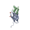
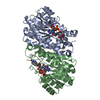
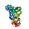
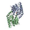
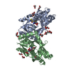
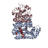
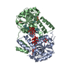
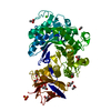
Crystal structure of the sixth cohesin from Acetivibrio cellulolyticus' scaffoldin B in complex with Cel5 dockerin S15I, I16N mutant
Components
  • DocCel5: Type I dockerin repeat domain from A. cellulolyticus family 5 endoglucanase WP_010249057 S15I, I16N mutant
  • Endoglucanase
KeywordsPROTEIN BINDING / cellulosome / cohesin / dockerin / type I cohesin-dockerin / Coh-Doc / protein-protein interaction
Function / homology
Function and homology information


cellulose binding / cellulase / cellulase activity / cellulose catabolic process / polysaccharide catabolic process / hydrolase activity, hydrolyzing O-glycosyl compounds / extracellular region / metal ion binding
Similarity search - Function
Type 1 dockerin domain / Dockerin domain / CarboxypepD_reg-like domain / Glycosyl hydrolases family 9, Asp/Glu active sites / Glycosyl hydrolases family 9 (GH9) active site signature 3. / Cellulosome anchoring protein, cohesin domain / Cohesin domain / Carboxypeptidase-like, regulatory domain superfamily / Cellulose binding domain / Cellulose binding domain ...Type 1 dockerin domain / Dockerin domain / CarboxypepD_reg-like domain / Glycosyl hydrolases family 9, Asp/Glu active sites / Glycosyl hydrolases family 9 (GH9) active site signature 3. / Cellulosome anchoring protein, cohesin domain / Cohesin domain / Carboxypeptidase-like, regulatory domain superfamily / Cellulose binding domain / Cellulose binding domain / Carbohydrate-binding module 3 / Carbohydrate-binding module 3 superfamily / CBM3 (carbohydrate binding type-3) domain profile. / Glycoside hydrolase family 9 / Glycosyl hydrolase family 9 / Dockerin domain / Dockerin domain profile. / Dockerin type I domain / Dockerin type I repeat / Dockerin domain superfamily / CBM2/CBM3, carbohydrate-binding domain superfamily / Six-hairpin glycosidase-like superfamily / Six-hairpin glycosidase superfamily / EF-Hand 1, calcium-binding site / EF-hand calcium-binding domain. / Orthogonal Bundle / Mainly Alpha
Similarity search - Domain/homology
THIOCYANATE ION / DocCel5: Type I dockerin repeat domain from A. cellulolyticus family 5 endoglucanase WP_010249057 S15I, I16N mutant / Endoglucanase
Similarity search - Component
Biological speciesAcetivibrio cellulolyticus (bacteria)
MethodX-RAY DIFFRACTION / SYNCHROTRON / MOLECULAR REPLACEMENT / Resolution: 1.45 Å
AuthorsBule, P. / Najmudin, S. / Fontes, C.M.G.A. / Alves, V.D.
Funding support Portugal, 3items
OrganizationGrant numberCountry
Fundacao para a Ciencia e TecnologiaEXPL/BIA-MIC/1176/2012 Portugal
Fundacao para a Ciencia e TecnologiaPTDC/BIA-PRO/103980/2008 Portugal
Fundacao para a Ciencia e TecnologiaPTDC/BIA-MIC/5947/2014 Portugal
CitationJournal: J. Biol. Chem. / Year: 2018
Title: Structure-function analyses generate novel specificities to assemble the components of multienzyme bacterial cellulosome complexes.
Authors: Bule, P. / Cameron, K. / Prates, J.A.M. / Ferreira, L.M.A. / Smith, S.P. / Gilbert, H.J. / Bayer, E.A. / Najmudin, S. / Fontes, C.M.G.A. / Alves, V.D.
History
DepositionApr 24, 2017Deposition site: PDBE / Processing site: PDBE
Revision 1.0Jan 31, 2018Provider: repository / Type: Initial release
Revision 1.1Feb 7, 2018Group: Database references / Category: citation / citation_author
Item: _citation.journal_abbrev / _citation.pdbx_database_id_DOI ..._citation.journal_abbrev / _citation.pdbx_database_id_DOI / _citation.pdbx_database_id_PubMed / _citation.title / _citation_author.name
Revision 1.2Apr 25, 2018Group: Data collection / Database references / Category: citation
Item: _citation.journal_volume / _citation.page_first ..._citation.journal_volume / _citation.page_first / _citation.page_last / _citation.title
Revision 1.3Jan 17, 2024Group: Data collection / Database references ...Data collection / Database references / Derived calculations / Refinement description
Category: chem_comp_atom / chem_comp_bond ...chem_comp_atom / chem_comp_bond / database_2 / pdbx_initial_refinement_model / struct_conn
Item: _database_2.pdbx_DOI / _database_2.pdbx_database_accession ..._database_2.pdbx_DOI / _database_2.pdbx_database_accession / _struct_conn.pdbx_dist_value / _struct_conn.ptnr1_auth_asym_id / _struct_conn.ptnr1_auth_comp_id / _struct_conn.ptnr1_auth_seq_id / _struct_conn.ptnr1_label_asym_id / _struct_conn.ptnr1_label_atom_id / _struct_conn.ptnr1_label_comp_id / _struct_conn.ptnr1_label_seq_id / _struct_conn.ptnr2_auth_asym_id / _struct_conn.ptnr2_auth_comp_id / _struct_conn.ptnr2_auth_seq_id / _struct_conn.ptnr2_label_asym_id / _struct_conn.ptnr2_label_atom_id / _struct_conn.ptnr2_label_comp_id

-
Structure visualization

Structure viewerMolecule:
MolmilJmol/JSmol

Downloads & links

-
Assembly

Deposited unit
A: Endoglucanase
B: DocCel5: Type I dockerin repeat domain from A. cellulolyticus family 5 endoglucanase WP_010249057 S15I, I16N mutant
C: Endoglucanase
D: DocCel5: Type I dockerin repeat domain from A. cellulolyticus family 5 endoglucanase WP_010249057 S15I, I16N mutant
hetero molecules


Theoretical massNumber of molelcules
Total (without water)45,07715
Polymers44,4784
Non-polymers59911
Water10,449580
1


  • Idetical with deposited unit
  • defined by author&software
  • Evidence: isothermal titration calorimetry
TypeNameSymmetry operationNumber
identity operation1_555x,y,z1
Buried area6020 Å2
ΔGint-82 kcal/mol
Surface area18070 Å2
MethodPISA
Unit cell
Length a, b, c (Å)46.539, 79.809, 112.159
Angle α, β, γ (deg.)90.000, 90.000, 90.000
Int Tables number19
Space group name H-MP212121

-
Components

-
Protein , 2 types, 4 molecules ACBD

#1: Protein Endoglucanase / scaffoldin


Mass: 14747.412 Da / Num. of mol.: 2 / Fragment: ScaB Type I cohesin domain UNP residues 1472-1611
Source method: isolated from a genetically manipulated source
Source: (gene. exp.) Acetivibrio cellulolyticus (bacteria) / Gene: cipV / Plasmid: pET28a / Production host: Escherichia coli BL21(DE3) (bacteria) / References: UniProt: Q9RPL0, cellulase
#2: Protein DocCel5: Type I dockerin repeat domain from A. cellulolyticus family 5 endoglucanase WP_010249057 S15I, I16N mutant


Mass: 7491.539 Da / Num. of mol.: 2 / Fragment: Type I dockerin domain
Source method: isolated from a genetically manipulated source
Source: (gene. exp.) Acetivibrio cellulolyticus (bacteria) / Gene: BglC / Plasmid: pET28a / Production host: Escherichia coli BL21(DE3) (bacteria) / References: UniProt: A0A2R2JFJ6*PLUS

-
Non-polymers , 4 types, 591 molecules

#3: Chemical
ChemComp-CA / CALCIUM ION


Mass: 40.078 Da / Num. of mol.: 6 / Source method: obtained synthetically / Formula: Ca
#4: Chemical ChemComp-SCN / THIOCYANATE ION


Mass: 58.082 Da / Num. of mol.: 3 / Source method: obtained synthetically / Formula: CNS
#5: Chemical ChemComp-GOL / GLYCEROL / GLYCERIN / PROPANE-1,2,3-TRIOL


Mass: 92.094 Da / Num. of mol.: 2 / Source method: obtained synthetically / Formula: C3H8O3
#6: Water ChemComp-HOH / water


Mass: 18.015 Da / Num. of mol.: 580 / Source method: isolated from a natural source / Formula: H2O

-
Experimental details

-
Experiment

ExperimentMethod: X-RAY DIFFRACTION / Number of used crystals: 1

-
Sample preparation

CrystalDensity Matthews: 2.34 Å3/Da / Density % sol: 47.47 % / Mosaicity: 0.18 °
Crystal growTemperature: 292 K / Method: vapor diffusion, hanging drop / pH: 6.9 / Details: 0.2 M sodium thiocyanate, 20% (w/v) PEG 3350

-
Data collection

DiffractionMean temperature: 100 K
Diffraction sourceSource: SYNCHROTRON / Site: ESRF / Beamline: ID29 / Wavelength: 0.9763 Å
DetectorType: DECTRIS PILATUS 6M-F / Detector: PIXEL / Date: Dec 13, 2013
RadiationProtocol: SINGLE WAVELENGTH / Monochromatic (M) / Laue (L): M / Scattering type: x-ray
Radiation wavelengthWavelength: 0.9763 Å / Relative weight: 1
ReflectionResolution: 1.45→27.38 Å / Num. obs: 74140 / % possible obs: 99.2 % / Redundancy: 4.4 % / CC1/2: 0.998 / Rmerge(I) obs: 0.057 / Rpim(I) all: 0.028 / Rrim(I) all: 0.064 / Net I/σ(I): 14.9 / Num. measured all: 327094 / Scaling rejects: 108
Reflection shell

Diffraction-ID: 1

Resolution (Å)Redundancy (%)Rmerge(I) obsNum. measured allNum. unique allCC1/2Rpim(I) allRrim(I) allNet I/σ(I) obs% possible all
1.45-1.472.60.203880733950.9120.1410.254.193.8
7.94-27.384.60.04124235230.9970.020.04630.297.5

-
Processing

Software
NameVersionClassification
REFMAC5.8.0073refinement
Aimless0.3.11data scaling
PDB_EXTRACT3.22data extraction
iMOSFLMdata reduction
PHASERphasing
RefinementMethod to determine structure: MOLECULAR REPLACEMENT
Starting model: 2ccl
Resolution: 1.45→65.03 Å / Cor.coef. Fo:Fc: 0.972 / Cor.coef. Fo:Fc free: 0.962 / SU B: 1.645 / SU ML: 0.034 / Cross valid method: THROUGHOUT / σ(F): 0 / ESU R: 0.057 / ESU R Free: 0.059 / Stereochemistry target values: MAXIMUM LIKELIHOOD
Details: HYDROGENS HAVE BEEN ADDED IN THE RIDING POSITIONS U VALUES : WITH TLS ADDED
RfactorNum. reflection% reflectionSelection details
Rfree0.1699 3641 4.9 %RANDOM
Rwork0.144 ---
obs0.1453 70443 98.98 %-
Solvent computationIon probe radii: 0.7 Å / Shrinkage radii: 0.7 Å / VDW probe radii: 1.2 Å / Solvent model: MASK
Displacement parametersBiso max: 67.86 Å2 / Biso mean: 14.99 Å2 / Biso min: 4.77 Å2
Baniso -1Baniso -2Baniso -3
1--0.22 Å20 Å2-0 Å2
2--0.19 Å20 Å2
3---0.03 Å2
Refinement stepCycle: final / Resolution: 1.45→65.03 Å
ProteinNucleic acidLigandSolventTotal
Num. atoms3114 0 27 580 3721
Biso mean--23.46 26.94 -
Num. residues----421
Refine LS restraints
Refine-IDTypeDev idealDev ideal targetNumber
X-RAY DIFFRACTIONr_bond_refined_d0.0090.023331
X-RAY DIFFRACTIONr_bond_other_d0.0050.023240
X-RAY DIFFRACTIONr_angle_refined_deg1.4961.9844527
X-RAY DIFFRACTIONr_angle_other_deg1.0937526
X-RAY DIFFRACTIONr_dihedral_angle_1_deg6.5275465
X-RAY DIFFRACTIONr_dihedral_angle_2_deg34.16826.585123
X-RAY DIFFRACTIONr_dihedral_angle_3_deg10.79315593
X-RAY DIFFRACTIONr_dihedral_angle_4_deg3.311154
X-RAY DIFFRACTIONr_chiral_restr0.0880.2539
X-RAY DIFFRACTIONr_gen_planes_refined0.0070.023820
X-RAY DIFFRACTIONr_gen_planes_other0.0050.02680
LS refinement shellResolution: 1.45→1.488 Å / Rfactor Rfree error: 0 / Total num. of bins used: 20
RfactorNum. reflection% reflection
Rfree0.221 269 -
Rwork0.183 4859 -
all-5128 -
obs--94.32 %
Refinement TLS params.

Method: refined / Refine-ID: X-RAY DIFFRACTION

IDL112)L122)L132)L222)L232)L332)S11 (Å °)S12 (Å °)S13 (Å °)S21 (Å °)S22 (Å °)S23 (Å °)S31 (Å °)S32 (Å °)S33 (Å °)T112)T122)T132)T222)T232)T332)Origin x (Å)Origin y (Å)Origin z (Å)
10.3974-0.2404-0.13770.43240.11320.7002-0.01490.0221-0.0074-0.0419-0.00890.00720.0111-0.02760.02380.025-0.0095-0.00070.0139-0.00780.007812.20673.083-48.846
22.0191-0.15970.2940.89710.37711.17150.0153-0.2251-0.13380.0948-0.00830.02450.088-0.092-0.0070.0364-0.01410.00280.05590.00280.01397.49875.371-25.918
30.6245-0.16030.27391.08390.29161.9425-0.0065-0.048-0.11070.1223-0.0130.02630.3867-0.00850.01940.11440.00410.01130.0289-0.00650.03330.28473.289-10.572
41.70310.35220.76080.8090.17722.2505-0.07990.16580.0747-0.10150.05120.0109-0.1250.13880.02880.0373-0.0083-0.00320.0542-0.01030.018427.98784.585-31.413
Refinement TLS group
IDRefine-IDRefine TLS-IDAuth asym-IDAuth seq-ID
1X-RAY DIFFRACTION1A0 - 142
2X-RAY DIFFRACTION2B3 - 70
3X-RAY DIFFRACTION3C0 - 142
4X-RAY DIFFRACTION4D3 - 69

+
About Yorodumi

-
News

-
Feb 9, 2022. New format data for meta-information of EMDB entries

New format data for meta-information of EMDB entries

  • Version 3 of the EMDB header file is now the official format.
  • The previous official version 1.9 will be removed from the archive.

Related info.:EMDB header

External links:wwPDB to switch to version 3 of the EMDB data model

-
Aug 12, 2020. Covid-19 info

Covid-19 info

URL: https://pdbjlvh1.pdbj.org/emnavi/covid19.php

New page: Covid-19 featured information page in EM Navigator.

Related info.:Covid-19 info / Mar 5, 2020. Novel coronavirus structure data

+
Mar 5, 2020. Novel coronavirus structure data

Novel coronavirus structure data

Related info.:Yorodumi Speices / Aug 12, 2020. Covid-19 info

External links:COVID-19 featured content - PDBj / Molecule of the Month (242):Coronavirus Proteases

+
Jan 31, 2019. EMDB accession codes are about to change! (news from PDBe EMDB page)

EMDB accession codes are about to change! (news from PDBe EMDB page)

  • The allocation of 4 digits for EMDB accession codes will soon come to an end. Whilst these codes will remain in use, new EMDB accession codes will include an additional digit and will expand incrementally as the available range of codes is exhausted. The current 4-digit format prefixed with “EMD-” (i.e. EMD-XXXX) will advance to a 5-digit format (i.e. EMD-XXXXX), and so on. It is currently estimated that the 4-digit codes will be depleted around Spring 2019, at which point the 5-digit format will come into force.
  • The EM Navigator/Yorodumi systems omit the EMD- prefix.

Related info.:Q: What is EMD? / ID/Accession-code notation in Yorodumi/EM Navigator

External links:EMDB Accession Codes are Changing Soon! / Contact to PDBj

+
Jul 12, 2017. Major update of PDB

Major update of PDB

  • wwPDB released updated PDB data conforming to the new PDBx/mmCIF dictionary.
  • This is a major update changing the version number from 4 to 5, and with Remediation, in which all the entries are updated.
  • In this update, many items about electron microscopy experimental information are reorganized (e.g. em_software).
  • Now, EM Navigator and Yorodumi are based on the updated data.

External links:wwPDB Remediation / Enriched Model Files Conforming to OneDep Data Standards Now Available in the PDB FTP Archive

-
Yorodumi

Thousand views of thousand structures

  • Yorodumi is a browser for structure data from EMDB, PDB, SASBDB, etc.
  • This page is also the successor to EM Navigator detail page, and also detail information page/front-end page for Omokage search.
  • The word "yorodu" (or yorozu) is an old Japanese word meaning "ten thousand". "mi" (miru) is to see.

Related info.:EMDB / PDB / SASBDB / Comparison of 3 databanks / Yorodumi Search / Aug 31, 2016. New EM Navigator & Yorodumi / Yorodumi Papers / Jmol/JSmol / Function and homology information / Changes in new EM Navigator and Yorodumi

Read more