 Movie
Movie Controller

[English] 日本語
 Yorodumi
- PDB-5h9d: Crystal structure of Heptaprenyl Diphosphate Synthase from Staphy... -
+ Open data

- Basic information
| Entry | Database: PDB / ID: 5h9d | ||||||
|---|---|---|---|---|---|---|---|
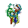
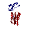
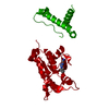
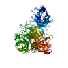
| Title | Crystal structure of Heptaprenyl Diphosphate Synthase from Staphylococcus aureus | ||||||
 Components Components |
| ||||||
 Keywords Keywords | TRANSFERASE / metal-binding / substrate binding / acidocalcisomal pyrophosphatase | ||||||
| Function / homology |  Function and homology information Function and homology informationheptaprenyl diphosphate synthase activity / heptaprenyl diphosphate synthase / dimethylallyltranstransferase / prenyltransferase activity / menaquinone biosynthetic process / isoprenoid biosynthetic process / dimethylallyltranstransferase activity Similarity search - Function | ||||||
| Biological species |   Staphylococcus aureus (bacteria) Staphylococcus aureus (bacteria) | ||||||
| Method |  X-RAY DIFFRACTION / SYNCHROTRON / MOLECULAR REPLACEMENT / molecular replacement / Resolution: 2.68 Å X-RAY DIFFRACTION / SYNCHROTRON / MOLECULAR REPLACEMENT / molecular replacement / Resolution: 2.68 Å | ||||||
 Authors Authors | Wei, H.L. / Liu, W.D. / Zheng, Y.Y. / Ko, T.P. / Chen, C.C. / Guo, R.T. | ||||||
 Citation Citation | Journal: ChemMedChem / Year: 2016 Title: Structure, Function, and Inhibition of Staphylococcus aureus Heptaprenyl Diphosphate Synthase Authors: Desai, J. / Liu, Y.L. / Wei, H. / Liu, W. / Ko, T.P. / Guo, R.T. / Oldfield, E. | ||||||
| History |
|
- Structure visualization
| Structure viewer | Molecule: MolmilJmol/JSmol |
|---|
- Downloads & links
-Download
| PDBx/mmCIF format | 5h9d.cif.gz | 218.4 KB | Display | PDBx/mmCIF format |
|---|---|---|---|---|
| PDB format | pdb5h9d.ent.gz | 176.6 KB | Display | PDB format |
| PDBx/mmJSON format | 5h9d.json.gz | Tree view | PDBx/mmJSON format | |
| Others |  Other downloads Other downloads |
-Validation report
| Arichive directory | https://data.pdbj.org/pub/pdb/validation_reports/h9/5h9dftp://data.pdbj.org/pub/pdb/validation_reports/h9/5h9d | HTTPS FTP |
|---|
-Related structure data
| Similar structure data |
|---|


-Links
 PDBj
PDBj
- Assembly
| Deposited unit | 
| |||||||||
|---|---|---|---|---|---|---|---|---|---|---|
| 1 | 
| |||||||||
| 2 | 
| |||||||||
| Unit cell |
| |||||||||
| Components on special symmetry positions |
|
-Components
| #1: Protein | Mass: 36199.906 Da / Num. of mol.: 2 Source method: isolated from a genetically manipulated source Source: (gene. exp.) Staphylococcus aureus (bacteria)Gene: hepT, gerCC, AFO87_08490, AL078_00755, AL493_08930, AL498_03950, AL508_07350, AM595_07380, CH51_07695, EQ80_006970, ER12_006960, ERS092844_00891, ERS093009_01199, ERS195423_01812, ERS365775_ ...Gene: hepT, gerCC, AFO87_08490, AL078_00755, AL493_08930, AL498_03950, AL508_07350, AM595_07380, CH51_07695, EQ80_006970, ER12_006960, ERS092844_00891, ERS093009_01199, ERS195423_01812, ERS365775_02304, ERS410449_02217, ERS411009_00914, ERS411017_01500, ERS445052_01888, FE68_02720, RL02_11030, RT87_07355, SAMI_1402 Plasmid: pET28 / Production host: References: UniProt: A0A0D6HKK2, UniProt: Q2FYG6*PLUS, dimethylallyltranstransferase, heptaprenyl diphosphate synthase #2: Protein | Mass: 22185.957 Da / Num. of mol.: 2 Source method: isolated from a genetically manipulated source Source: (gene. exp.) Staphylococcus aureus (bacteria)Gene: AL078_00765, AM595_07390, AUC48_07185, AUC49_07360, AUC50_07510, BN1321_260110, ERS179246_00728, ERS445051_01360, NI36_07270, RT87_07365, SA7112_09025, SAFDA_1366, SAMI_1404, SASCBU26_01375 Plasmid: pET28 / Production host: #3: Protein/peptide | Mass: 2093.390 Da / Num. of mol.: 2 Source method: isolated from a genetically manipulated source Source: (gene. exp.) Staphylococcus aureus (bacteria) / Plasmid: pET28 / Production host: #4: Chemical | ChemComp-PO4 /   Mass: 94.971 Da / Num. of mol.: 4 / Source method: obtained synthetically / Formula: PO4 Mass: 94.971 Da / Num. of mol.: 4 / Source method: obtained synthetically / Formula: PO4#5: Water | ChemComp-HOH / |  Mass: 18.015 Da / Num. of mol.: 118 / Source method: isolated from a natural source / Formula: H2O Mass: 18.015 Da / Num. of mol.: 118 / Source method: isolated from a natural source / Formula: H2O |
|---|
-Experimental details
-Experiment
| Experiment | Method: X-RAY DIFFRACTION / Number of used crystals: 1 |
|---|
- Sample preparation
| Crystal | Density Matthews: 2.92 Å3/Da / Density % sol: 57.82 % |
|---|---|
| Crystal grow | Temperature: 298 K / Method: vapor diffusion, sitting drop / pH: 7.6 / Details: Citic acid, bis-tris propane, PEG 3350 |
-Data collection
| Diffraction | Mean temperature: 100 K | |||||||||||||||||||||||||||||||||||||||||||||||||||||||||||||||||||||||||||||||||||||||||||||||||||
|---|---|---|---|---|---|---|---|---|---|---|---|---|---|---|---|---|---|---|---|---|---|---|---|---|---|---|---|---|---|---|---|---|---|---|---|---|---|---|---|---|---|---|---|---|---|---|---|---|---|---|---|---|---|---|---|---|---|---|---|---|---|---|---|---|---|---|---|---|---|---|---|---|---|---|---|---|---|---|---|---|---|---|---|---|---|---|---|---|---|---|---|---|---|---|---|---|---|---|---|---|
| Diffraction source | Source: SYNCHROTRON / Site: NSRRC  / Beamline: BL13B1 / Wavelength: 1 Å / Beamline: BL13B1 / Wavelength: 1 Å | |||||||||||||||||||||||||||||||||||||||||||||||||||||||||||||||||||||||||||||||||||||||||||||||||||
| Detector | Type: ADSC QUANTUM 315r / Detector: CCD / Date: Apr 9, 2015 | |||||||||||||||||||||||||||||||||||||||||||||||||||||||||||||||||||||||||||||||||||||||||||||||||||
| Radiation | Monochromator: GRAPHITE / Protocol: SINGLE WAVELENGTH / Monochromatic (M) / Laue (L): M / Scattering type: x-ray | |||||||||||||||||||||||||||||||||||||||||||||||||||||||||||||||||||||||||||||||||||||||||||||||||||
| Radiation wavelength | Wavelength: 1 Å / Relative weight: 1 | |||||||||||||||||||||||||||||||||||||||||||||||||||||||||||||||||||||||||||||||||||||||||||||||||||
| Reflection | Resolution: 2.68→25 Å / Num. obs: 35313 / % possible obs: 92 % / Redundancy: 5 % / Rmerge(I) obs: 0.052 / Rpim(I) all: 0.025 / Rrim(I) all: 0.057 / Χ2: 2.258 / Net I/av σ(I): 38.038 / Net I/σ(I): 25 / Num. measured all: 175144 | |||||||||||||||||||||||||||||||||||||||||||||||||||||||||||||||||||||||||||||||||||||||||||||||||||
| Reflection shell | Diffraction-ID: 1 / Rejects: _
|
-Phasing
| Phasing | Method: molecular replacement |
|---|
- Processing
| Software |
| ||||||||||||||||||||
|---|---|---|---|---|---|---|---|---|---|---|---|---|---|---|---|---|---|---|---|---|---|
| Refinement | Method to determine structure: MOLECULAR REPLACEMENT / Resolution: 2.68→25 Å / Cross valid method: FREE R-VALUE / σ(F): 0 Details: Chains K/L are built from residual densities exist between the neighboring heterodimers. Author persume Chains K/L come from a neighboring heterodimer with the region 140-170 of the protein unfolded.
| ||||||||||||||||||||
| Solvent computation | Bsol: 50.3455 Å2 | ||||||||||||||||||||
| Displacement parameters | Biso max: 148.41 Å2 / Biso mean: 75.7839 Å2 / Biso min: 37.28 Å2
| ||||||||||||||||||||
| Refinement step | Cycle: LAST / Resolution: 2.68→25 Å
| ||||||||||||||||||||
| Refine LS restraints |
|
