 Movie
Movie Controller

[English] 日本語
 Yorodumi
- PDB-5npy: Crystal structure of Helicobacter pylori flagellar hook protein FlgE2 -
+ Open data

- Basic information
| Entry | Database: PDB / ID: 5npy | ||||||
|---|---|---|---|---|---|---|---|
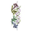
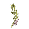
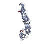
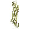
| Title | Crystal structure of Helicobacter pylori flagellar hook protein FlgE2 | ||||||
 Components Components | Flagellar basal body protein | ||||||
 Keywords Keywords | MOTOR PROTEIN / Helicobactyer pylori / flagellum / hook | ||||||
| Function / homology |  Function and homology information Function and homology informationbacterial-type flagellum basal body / bacterial-type flagellum-dependent cell motility Similarity search - Function | ||||||
| Biological species |   Helicobacter pylori (bacteria) Helicobacter pylori (bacteria) | ||||||
| Method |  X-RAY DIFFRACTION / SYNCHROTRON / SAD / Resolution: 2.292 Å X-RAY DIFFRACTION / SYNCHROTRON / SAD / Resolution: 2.292 Å | ||||||
 Authors Authors | Loconte, V. / Zanotti, G. / Kekez, I. / Matkovic-Calogovic, D. | ||||||
 Citation Citation | Journal: FEBS J. / Year: 2017 Title: Structural characterization of FlgE2 protein from Helicobacter pylori hook. Authors: Loconte, V. / Kekez, I. / Matkovic-Calogovic, D. / Zanotti, G. | ||||||
| History |
|
- Structure visualization
| Structure viewer | Molecule: MolmilJmol/JSmol |
|---|
- Downloads & links
-Download
| PDBx/mmCIF format | 5npy.cif.gz | 214.6 KB | Display | PDBx/mmCIF format |
|---|---|---|---|---|
| PDB format | pdb5npy.ent.gz | 169.8 KB | Display | PDB format |
| PDBx/mmJSON format | 5npy.json.gz | Tree view | PDBx/mmJSON format | |
| Others |  Other downloads Other downloads |
-Validation report
| Arichive directory | https://data.pdbj.org/pub/pdb/validation_reports/np/5npyftp://data.pdbj.org/pub/pdb/validation_reports/np/5npy | HTTPS FTP |
|---|
-Related structure data
| Similar structure data |
|---|


-Links
 PDBj
PDBj
- Assembly
| Deposited unit | 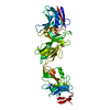
| ||||||||
|---|---|---|---|---|---|---|---|---|---|
| 1 |
| ||||||||
| Unit cell |
|
-Components
| #1: Protein | Mass: 68904.969 Da / Num. of mol.: 1 Source method: isolated from a genetically manipulated source Source: (gene. exp.) Helicobacter pylori (bacteria) / Strain: G27 / Gene: flgE-2, HPG27_859 / Plasmid: pETit-hp0908 / Production host: |
|---|---|
| #2: Chemical | ChemComp-144 /   Mass: 122.143 Da / Num. of mol.: 1 / Source method: obtained synthetically / Formula: C4H12NO3 Mass: 122.143 Da / Num. of mol.: 1 / Source method: obtained synthetically / Formula: C4H12NO3 |
| #3: Water | ChemComp-HOH /  Mass: 18.015 Da / Num. of mol.: 308 / Source method: isolated from a natural source / Formula: H2O Mass: 18.015 Da / Num. of mol.: 308 / Source method: isolated from a natural source / Formula: H2O |
-Experimental details
-Experiment
| Experiment | Method: X-RAY DIFFRACTION / Number of used crystals: 1 |
|---|
- Sample preparation
| Crystal | Density Matthews: 2.32 Å3/Da / Density % sol: 47 % |
|---|---|
| Crystal grow | Temperature: 293 K / Method: vapor diffusion, sitting drop / pH: 8.5 Details: 0.2 M Potassium Thiocyanate, 0.1 M Bis-Tris Propane pH 8.5, 20% w/v PEG3350 |
-Data collection
| Diffraction |
| |||||||||||||||
|---|---|---|---|---|---|---|---|---|---|---|---|---|---|---|---|---|
| Diffraction source |
| |||||||||||||||
| Detector |
| |||||||||||||||
| Radiation |
| |||||||||||||||
| Radiation wavelength |
| |||||||||||||||
| Reflection | Resolution: 2.29→48.551 Å / Num. obs: 23979 / % possible obs: 98.5 % / Redundancy: 5.5 % / Rmerge(I) obs: 0.148 / Rpim(I) all: 0.076 / Net I/σ(I): 9.7 | |||||||||||||||
| Reflection shell | Resolution: 2.29→2.4 Å / Redundancy: 4.8 % / Rmerge(I) obs: 0.671 / Mean I/σ(I) obs: 2.3 / Num. unique obs: 3106 / Rpim(I) all: 0.362 / % possible all: 90.8 |

- Processing
| Software |
| ||||||||||||||||||||||||||||||||||||||||||||||||||||||||||||||||||||||||||||||||||||||||||||||||||||||||||||||||
|---|---|---|---|---|---|---|---|---|---|---|---|---|---|---|---|---|---|---|---|---|---|---|---|---|---|---|---|---|---|---|---|---|---|---|---|---|---|---|---|---|---|---|---|---|---|---|---|---|---|---|---|---|---|---|---|---|---|---|---|---|---|---|---|---|---|---|---|---|---|---|---|---|---|---|---|---|---|---|---|---|---|---|---|---|---|---|---|---|---|---|---|---|---|---|---|---|---|---|---|---|---|---|---|---|---|---|---|---|---|---|---|---|---|
| Refinement | Method to determine structure: SAD / Resolution: 2.292→48.551 Å / SU ML: 0.35 / Data cutoff high absF: 0 / Data cutoff low absF: 0 / Cross valid method: FREE R-VALUE / σ(F): 1.25 / Phase error: 28.21
| ||||||||||||||||||||||||||||||||||||||||||||||||||||||||||||||||||||||||||||||||||||||||||||||||||||||||||||||||
| Solvent computation | Shrinkage radii: 0.9 Å / VDW probe radii: 1.11 Å | ||||||||||||||||||||||||||||||||||||||||||||||||||||||||||||||||||||||||||||||||||||||||||||||||||||||||||||||||
| Refinement step | Cycle: LAST / Resolution: 2.292→48.551 Å
| ||||||||||||||||||||||||||||||||||||||||||||||||||||||||||||||||||||||||||||||||||||||||||||||||||||||||||||||||
| Refine LS restraints |
| ||||||||||||||||||||||||||||||||||||||||||||||||||||||||||||||||||||||||||||||||||||||||||||||||||||||||||||||||
| LS refinement shell |
| ||||||||||||||||||||||||||||||||||||||||||||||||||||||||||||||||||||||||||||||||||||||||||||||||||||||||||||||||
| Refinement TLS params. | Method: refined / Origin x: 36.8348 Å / Origin y: -0.1189 Å / Origin z: 227.6481 Å
| ||||||||||||||||||||||||||||||||||||||||||||||||||||||||||||||||||||||||||||||||||||||||||||||||||||||||||||||||
| Refinement TLS group | Selection details: all |
