 Movie
Movie Controller


+ Open data

- Basic information
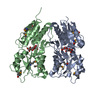
| Entry | Database: PDB / ID: 5npu | ||||||
|---|---|---|---|---|---|---|---|
| Title | Inferred ancestral pyruvate decarboxylase | ||||||
 Components Components | ANC27 | ||||||
 Keywords Keywords | LYASE / PYRUVATE DECARBOXYLASE / Ancestral Sequence Reconstruction | ||||||
| Function / homology | Thiamin diphosphate (ThDP)-binding fold, Pyr/PP domains / TPP-binding domain / Rossmann fold / 3-Layer(aba) Sandwich / Alpha Beta / DI(HYDROXYETHYL)ETHER / THIAMINE DIPHOSPHATE Function and homology information Function and homology information | ||||||
| Biological species | synthetic construct (others) | ||||||
| Method |  X-RAY DIFFRACTION / SYNCHROTRON / MOLECULAR REPLACEMENT / Resolution: 3.5 Å X-RAY DIFFRACTION / SYNCHROTRON / MOLECULAR REPLACEMENT / Resolution: 3.5 Å | ||||||
 Authors Authors | Buddrus, L. / Crennell, S.J. / Leak, D.J. / Danson, M.J. / Andrews, E.S.V. / Arcus, V.L. | ||||||
 Citation Citation | Journal: Acta Crystallogr F Struct Biol Commun / Year: 2018 Title: Crystal structure of an inferred ancestral bacterial pyruvate decarboxylase. Authors: Buddrus, L. / Andrews, E.S.V. / Leak, D.J. / Danson, M.J. / Arcus, V.L. / Crennell, S.J. | ||||||
| History |
|
- Structure visualization
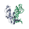
| Structure viewer | Molecule: MolmilJmol/JSmol |
|---|
- Downloads & links
-Download
| PDBx/mmCIF format | 5npu.cif.gz | 1.2 MB | Display | PDBx/mmCIF format |
|---|---|---|---|---|
| PDB format | pdb5npu.ent.gz | 1023.4 KB | Display | PDB format |
| PDBx/mmJSON format | 5npu.json.gz | Tree view | PDBx/mmJSON format | |
| Others |  Other downloads Other downloads |
-Validation report
| Arichive directory | https://data.pdbj.org/pub/pdb/validation_reports/np/5npuftp://data.pdbj.org/pub/pdb/validation_reports/np/5npu | HTTPS FTP |
|---|
-Related structure data
| Related structure data | 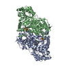 2wvgS S: Starting model for refinement |
|---|---|
| Similar structure data |


-Links
 PDBj
PDBj

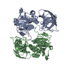
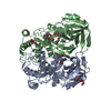
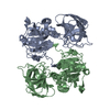
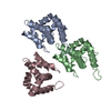
- Assembly
| Deposited unit | 
| ||||||||
|---|---|---|---|---|---|---|---|---|---|
| 1 | 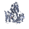
| ||||||||
| 2 | 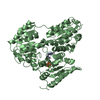
| ||||||||
| 3 | 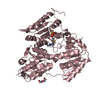
| ||||||||
| 4 | 
| ||||||||
| Unit cell |
|
-Components
| #1: Protein | Mass: 60700.902 Da / Num. of mol.: 4 Source method: isolated from a genetically manipulated source Source: (gene. exp.) synthetic construct (others) / Production host:  #2: Chemical | ChemComp-MG /   Mass: 24.305 Da / Num. of mol.: 4 / Source method: obtained synthetically / Formula: Mg Mass: 24.305 Da / Num. of mol.: 4 / Source method: obtained synthetically / Formula: Mg#3: Chemical | ChemComp-TPP /   Mass: 425.314 Da / Num. of mol.: 4 / Source method: obtained synthetically / Formula: C12H19N4O7P2S Mass: 425.314 Da / Num. of mol.: 4 / Source method: obtained synthetically / Formula: C12H19N4O7P2S#4: Chemical | ChemComp-PEG / |   Mass: 106.120 Da / Num. of mol.: 1 / Source method: isolated from a natural source / Formula: C4H10O3 Mass: 106.120 Da / Num. of mol.: 1 / Source method: isolated from a natural source / Formula: C4H10O3#5: Water | ChemComp-HOH / |  Mass: 18.015 Da / Num. of mol.: 1 / Source method: isolated from a natural source / Formula: H2O Mass: 18.015 Da / Num. of mol.: 1 / Source method: isolated from a natural source / Formula: H2O |
|---|
-Experimental details
-Experiment
| Experiment | Method: X-RAY DIFFRACTION / Number of used crystals: 1 |
|---|
- Sample preparation
| Crystal | Density Matthews: 2.25 Å3/Da / Density % sol: 45.36 % |
|---|---|
| Crystal grow | Temperature: 291 K / Method: vapor diffusion, hanging drop / Details: 0.15 M sodium citrate pH 5.5, 14%(w/v) PEG 3350 |
-Data collection
| Diffraction | Mean temperature: 100 K |
|---|---|
| Diffraction source | Source: SYNCHROTRON / Site: Australian Synchrotron  / Beamline: MX2 / Wavelength: 0.9537 Å / Beamline: MX2 / Wavelength: 0.9537 Å |
| Detector | Type: ADSC QUANTUM 315r / Detector: CCD / Date: Feb 26, 2016 / Details: FOCUSSING MIRRORS (TWO |
| Radiation | Monochromator: SI111 DOUBLE CRYSTAL / Protocol: SINGLE WAVELENGTH / Monochromatic (M) / Laue (L): M / Scattering type: x-ray |
| Radiation wavelength | Wavelength: 0.9537 Å / Relative weight: 1 |
| Reflection | Resolution: 3.5→93.82 Å / Num. obs: 28526 / % possible obs: 99.5 % / Redundancy: 4.4 % / Biso Wilson estimate: 53.8 Å2 / CC1/2: 0.965 / Rmerge(I) obs: 0.241 / Net I/σ(I): 5.6 |
| Reflection shell | Resolution: 3.5→3.71 Å / Redundancy: 4.6 % / Rmerge(I) obs: 0.965 / Mean I/σ(I) obs: 1.5 / Num. unique obs: 4512 / CC1/2: 0.422 / % possible all: 99.9 |
- Processing
| Software |
| |||||||||||||||||||||||||||||||||||||||||||||||||||||||||||||||||||||||||||||
|---|---|---|---|---|---|---|---|---|---|---|---|---|---|---|---|---|---|---|---|---|---|---|---|---|---|---|---|---|---|---|---|---|---|---|---|---|---|---|---|---|---|---|---|---|---|---|---|---|---|---|---|---|---|---|---|---|---|---|---|---|---|---|---|---|---|---|---|---|---|---|---|---|---|---|---|---|---|---|
| Refinement | Method to determine structure: MOLECULAR REPLACEMENT Starting model: 2wvg Resolution: 3.5→81.1 Å / SU ML: 0.7 / Cross valid method: THROUGHOUT / σ(F): 1.33 / Phase error: 36.18
| |||||||||||||||||||||||||||||||||||||||||||||||||||||||||||||||||||||||||||||
| Solvent computation | Shrinkage radii: 0.9 Å / VDW probe radii: 1.11 Å | |||||||||||||||||||||||||||||||||||||||||||||||||||||||||||||||||||||||||||||
| Refinement step | Cycle: LAST / Resolution: 3.5→81.1 Å
| |||||||||||||||||||||||||||||||||||||||||||||||||||||||||||||||||||||||||||||
| Refine LS restraints |
| |||||||||||||||||||||||||||||||||||||||||||||||||||||||||||||||||||||||||||||
| LS refinement shell |
| |||||||||||||||||||||||||||||||||||||||||||||||||||||||||||||||||||||||||||||
| Refinement TLS params. | Method: refined / Origin x: -1.4663 Å / Origin y: -51.0351 Å / Origin z: -27.8581 Å
| |||||||||||||||||||||||||||||||||||||||||||||||||||||||||||||||||||||||||||||
| Refinement TLS group | Selection details: all |
