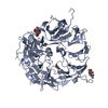
 Movie
Movie Controller


+ Open data

- Basic information
| Entry | Database: PDB / ID: 5nmr | |||||||||
|---|---|---|---|---|---|---|---|---|---|---|
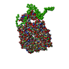
| Title | Monomeric mouse Sortilin extracellular domain | |||||||||
 Components Components | Sortilin | |||||||||
 Keywords Keywords | PROTEIN TRANSPORT / VPS10 domain family / sorting receptor / ten-bladed beta propeller / 10CC domain | |||||||||
| Function / homology |  Function and homology information Function and homology informationneurotensin receptor activity, non-G protein-coupled / nerve growth factor receptor activity / myotube differentiation / maintenance of synapse structure / cerebellar climbing fiber to Purkinje cell synapse / retromer complex binding / Golgi to lysosome transport / endosome transport via multivesicular body sorting pathway / Golgi Associated Vesicle Biogenesis / plasma membrane to endosome transport ...neurotensin receptor activity, non-G protein-coupled / nerve growth factor receptor activity / myotube differentiation / maintenance of synapse structure / cerebellar climbing fiber to Purkinje cell synapse / retromer complex binding / Golgi to lysosome transport / endosome transport via multivesicular body sorting pathway / Golgi Associated Vesicle Biogenesis / plasma membrane to endosome transport / nerve growth factor binding / vesicle organization / Golgi to endosome transport / trans-Golgi network transport vesicle / protein targeting to lysosome / positive regulation of epithelial cell apoptotic process / clathrin-coated vesicle / : / Golgi cisterna membrane / negative regulation of fat cell differentiation / endosome to lysosome transport / neurotrophin TRK receptor signaling pathway / extrinsic apoptotic signaling pathway via death domain receptors / clathrin-coated pit / ossification / cytoplasmic vesicle membrane / neuropeptide signaling pathway / response to insulin / endocytosis / regulation of gene expression / cytoplasmic vesicle / nuclear membrane / early endosome / lysosome / endosome membrane / G protein-coupled receptor signaling pathway / lysosomal membrane / neuronal cell body / dendrite / endoplasmic reticulum membrane / perinuclear region of cytoplasm / enzyme binding / cell surface / Golgi apparatus / membrane / plasma membrane / cytosol Similarity search - Function | |||||||||
| Biological species |  | |||||||||
| Method |  X-RAY DIFFRACTION / SYNCHROTRON / MOLECULAR REPLACEMENT / Resolution: 2.1 Å X-RAY DIFFRACTION / SYNCHROTRON / MOLECULAR REPLACEMENT / Resolution: 2.1 Å | |||||||||
 Authors Authors | Leloup, N.O.L. / Loessl, P. / Meijer, D.H.M. / Heck, A.J.R. / Thies-Weesie, D.M.E. / Janssen, B.J.C. | |||||||||
| Funding support |  Netherlands, 2items Netherlands, 2items
| |||||||||
 Citation Citation | Journal: Nat Commun / Year: 2017 Title: Low pH-induced conformational change and dimerization of sortilin triggers endocytosed ligand release. Authors: Nadia Leloup / Philip Lössl / Dimphna H Meijer / Martha Brennich / Albert J R Heck / Dominique M E Thies-Weesie / Bert J C Janssen /  Abstract: Low pH-induced ligand release and receptor recycling are important steps for endocytosis. The transmembrane protein sortilin, a β-propeller containing endocytosis receptor, internalizes a diverse ...Low pH-induced ligand release and receptor recycling are important steps for endocytosis. The transmembrane protein sortilin, a β-propeller containing endocytosis receptor, internalizes a diverse set of ligands with roles in cell differentiation and homeostasis. The molecular mechanisms of pH-mediated ligand release and sortilin recycling are unresolved. Here we present crystal structures that show the sortilin luminal segment (s-sortilin) undergoes a conformational change and dimerizes at low pH. The conformational change, within all three sortilin luminal domains, provides an altered surface and the dimers sterically shield a large interface while bringing the two s-sortilin C-termini into close proximity. Biophysical and cell-based assays show that members of two different ligand families, (pro)neurotrophins and neurotensin, preferentially bind the sortilin monomer. This indicates that sortilin dimerization and conformational change discharges ligands and triggers recycling. More generally, this work may reveal a double mechanism for low pH-induced ligand release by endocytosis receptors. | |||||||||
| History |
|
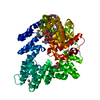
- Structure visualization
| Structure viewer | Molecule: MolmilJmol/JSmol |
|---|
- Downloads & links
-Download
| PDBx/mmCIF format | 5nmr.cif.gz | 256.6 KB | Display | PDBx/mmCIF format |
|---|---|---|---|---|
| PDB format | pdb5nmr.ent.gz | 204.1 KB | Display | PDB format |
| PDBx/mmJSON format | 5nmr.json.gz | Tree view | PDBx/mmJSON format | |
| Others |  Other downloads Other downloads |
-Validation report
| Arichive directory | https://data.pdbj.org/pub/pdb/validation_reports/nm/5nmrftp://data.pdbj.org/pub/pdb/validation_reports/nm/5nmr | HTTPS FTP |
|---|
-Related structure data
| Related structure data |  5nmtC  5nniC  5nnjC 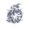 3f6kS S: Starting model for refinement C: citing same article ( |
|---|---|
| Similar structure data |


-Links
 PDBj
PDBj
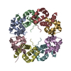
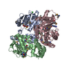
- Assembly
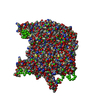
| Deposited unit | 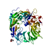
| ||||||||
|---|---|---|---|---|---|---|---|---|---|
| 1 |
| ||||||||
| Unit cell |
|
-Components
| #1: Protein | Mass: 81307.023 Da / Num. of mol.: 1 Source method: isolated from a genetically manipulated source Source: (gene. exp.)  Homo sapiens (human) / References: UniProt: Q6PHU5 Homo sapiens (human) / References: UniProt: Q6PHU5 | ||||||||
|---|---|---|---|---|---|---|---|---|---|
| #2: Polysaccharide | Source method: isolated from a genetically manipulated source #3: Sugar | ChemComp-NAG / |   Type: D-saccharide, beta linking / Mass: 221.208 Da / Num. of mol.: 1 Type: D-saccharide, beta linking / Mass: 221.208 Da / Num. of mol.: 1Source method: isolated from a genetically manipulated source Formula: C8H15NO6 #4: Chemical |   Mass: 40.078 Da / Num. of mol.: 2 / Source method: isolated from a natural source / Formula: Ca Mass: 40.078 Da / Num. of mol.: 2 / Source method: isolated from a natural source / Formula: Ca#5: Water | ChemComp-HOH / |  Mass: 18.015 Da / Num. of mol.: 226 / Source method: isolated from a natural source / Formula: H2O Mass: 18.015 Da / Num. of mol.: 226 / Source method: isolated from a natural source / Formula: H2OHas protein modification | Y | |
-Experimental details
-Experiment
| Experiment | Method: X-RAY DIFFRACTION / Number of used crystals: 1 |
|---|
- Sample preparation
| Crystal | Density Matthews: 2.66 Å3/Da / Density % sol: 53.74 % |
|---|---|
| Crystal grow | Temperature: 291 K / Method: vapor diffusion, sitting drop / pH: 7.5 Details: 0.1 M Na HEPES pH 7.5, 1 mM CaCl2 and 25% PEG 2000 MME (w/v). |
-Data collection
| Diffraction | Mean temperature: 100 K |
|---|---|
| Diffraction source | Source: SYNCHROTRON / Site: SLS  / Beamline: X06SA / Wavelength: 0.99998 Å / Beamline: X06SA / Wavelength: 0.99998 Å |
| Detector | Type: DECTRIS PILATUS3 6M / Detector: PIXEL / Date: Aug 29, 2014 |
| Radiation | Protocol: SINGLE WAVELENGTH / Monochromatic (M) / Laue (L): M / Scattering type: x-ray |
| Radiation wavelength | Wavelength: 0.99998 Å / Relative weight: 1 |
| Reflection | Resolution: 2→66 Å / Num. obs: 46680 / % possible obs: 97.5 % / Redundancy: 3.1 % / CC1/2: 0.985 / Rpim(I) all: 0.09 / Rsym value: 0.11 / Net I/σ(I): 6.3 |
- Processing
| Software |
| |||||||||||||||||||||||||||||||||||||||||||||||||||||||||||||||||||||||||||||||||||||||||||||||||||||||||||||||||||||||
|---|---|---|---|---|---|---|---|---|---|---|---|---|---|---|---|---|---|---|---|---|---|---|---|---|---|---|---|---|---|---|---|---|---|---|---|---|---|---|---|---|---|---|---|---|---|---|---|---|---|---|---|---|---|---|---|---|---|---|---|---|---|---|---|---|---|---|---|---|---|---|---|---|---|---|---|---|---|---|---|---|---|---|---|---|---|---|---|---|---|---|---|---|---|---|---|---|---|---|---|---|---|---|---|---|---|---|---|---|---|---|---|---|---|---|---|---|---|---|---|---|
| Refinement | Method to determine structure: MOLECULAR REPLACEMENT Starting model: 3F6K Resolution: 2.1→65.994 Å / SU ML: 0.27 / Cross valid method: FREE R-VALUE / σ(F): 1.34 / Phase error: 24.43 / Stereochemistry target values: ML
| |||||||||||||||||||||||||||||||||||||||||||||||||||||||||||||||||||||||||||||||||||||||||||||||||||||||||||||||||||||||
| Solvent computation | Shrinkage radii: 0.9 Å / VDW probe radii: 1.11 Å / Solvent model: FLAT BULK SOLVENT MODEL | |||||||||||||||||||||||||||||||||||||||||||||||||||||||||||||||||||||||||||||||||||||||||||||||||||||||||||||||||||||||
| Refinement step | Cycle: LAST / Resolution: 2.1→65.994 Å
| |||||||||||||||||||||||||||||||||||||||||||||||||||||||||||||||||||||||||||||||||||||||||||||||||||||||||||||||||||||||
| Refine LS restraints |
| |||||||||||||||||||||||||||||||||||||||||||||||||||||||||||||||||||||||||||||||||||||||||||||||||||||||||||||||||||||||
| LS refinement shell |
| |||||||||||||||||||||||||||||||||||||||||||||||||||||||||||||||||||||||||||||||||||||||||||||||||||||||||||||||||||||||
| Refinement TLS params. | Method: refined / Refine-ID: X-RAY DIFFRACTION
| |||||||||||||||||||||||||||||||||||||||||||||||||||||||||||||||||||||||||||||||||||||||||||||||||||||||||||||||||||||||
| Refinement TLS group |
|
