 Movie
Movie Controller

[English] 日本語
 Yorodumi
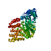
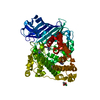
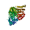
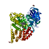
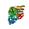
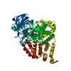
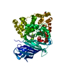
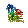
- PDB-5nia: Crystal structure of human LTA4H mutant D375N in open conformatio... -
+ Open data

- Basic information
| Entry | Database: PDB / ID: 5nia | ||||||
|---|---|---|---|---|---|---|---|
| Title | Crystal structure of human LTA4H mutant D375N in open conformation (crystal form I) | ||||||
 Components Components | Leukotriene A-4 hydrolase | ||||||
 Keywords Keywords | HYDROLASE / Metallopeptidase / epoxide hydrolase / open conformation | ||||||
| Function / homology |  Function and homology information Function and homology informationleukotriene-A4 hydrolase / leukotriene-A4 hydrolase activity / tripeptide aminopeptidase activity / tripeptide aminopeptidase / Biosynthesis of protectins / Biosynthesis of aspirin-triggered D-series resolvins / Biosynthesis of E-series 18(R)-resolvins / Biosynthesis of D-series resolvins / Biosynthesis of E-series 18(S)-resolvins / Synthesis of Leukotrienes (LT) and Eoxins (EX) ...leukotriene-A4 hydrolase / leukotriene-A4 hydrolase activity / tripeptide aminopeptidase activity / tripeptide aminopeptidase / Biosynthesis of protectins / Biosynthesis of aspirin-triggered D-series resolvins / Biosynthesis of E-series 18(R)-resolvins / Biosynthesis of D-series resolvins / Biosynthesis of E-series 18(S)-resolvins / Synthesis of Leukotrienes (LT) and Eoxins (EX) / epoxide hydrolase activity / leukotriene biosynthetic process / response to zinc ion / peptide catabolic process / metalloaminopeptidase activity / type I pneumocyte differentiation / aminopeptidase activity / lipid metabolic process / response to peptide hormone / tertiary granule lumen / peptidase activity / ficolin-1-rich granule lumen / Neutrophil degranulation / proteolysis / RNA binding / extracellular exosome / extracellular region / zinc ion binding / nucleoplasm / nucleus / cytosol Similarity search - Function | ||||||
| Biological species |  Homo sapiens (human) Homo sapiens (human) | ||||||
| Method |  X-RAY DIFFRACTION / SYNCHROTRON / MOLECULAR REPLACEMENT / Resolution: 1.764 Å X-RAY DIFFRACTION / SYNCHROTRON / MOLECULAR REPLACEMENT / Resolution: 1.764 Å | ||||||
 Authors Authors | Stsiapanava, A. | ||||||
| Funding support |  Sweden, 1items Sweden, 1items
| ||||||
 Citation Citation | Journal: Proc. Natl. Acad. Sci. U.S.A. / Year: 2017 Title: Capturing LTA4 hydrolase in action: Insights to the chemistry and dynamics of chemotactic LTB4 synthesis. Authors: Stsiapanava, A. / Samuelsson, B. / Haeggstrom, J.Z. | ||||||
| History |
|
- Structure visualization
| Structure viewer | Molecule: MolmilJmol/JSmol |
|---|
- Downloads & links
-Download
| PDBx/mmCIF format | 5nia.cif.gz | 375 KB | Display | PDBx/mmCIF format |
|---|---|---|---|---|
| PDB format | pdb5nia.ent.gz | 310.3 KB | Display | PDB format |
| PDBx/mmJSON format | 5nia.json.gz | Tree view | PDBx/mmJSON format | |
| Others |  Other downloads Other downloads |
-Validation report
| Arichive directory | https://data.pdbj.org/pub/pdb/validation_reports/ni/5niaftp://data.pdbj.org/pub/pdb/validation_reports/ni/5nia | HTTPS FTP |
|---|
-Related structure data
| Related structure data |  4dprC 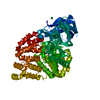 5ni2SC  5ni4C  5ni6C  5nidC  5nieC C: citing same article ( S: Starting model for refinement |
|---|---|
| Similar structure data |


-Links
 PDBj
PDBj- Assembly
| Deposited unit | 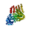
| ||||||||
|---|---|---|---|---|---|---|---|---|---|
| 1 |
| ||||||||
| Unit cell |
|
-Components
| #1: Protein | Mass: 70191.867 Da / Num. of mol.: 1 / Mutation: D375N Source method: isolated from a genetically manipulated source Source: (gene. exp.) Homo sapiens (human) / Gene: LTA4H, LTA4 / Plasmid: pT3MB4 / Production host:  |
|---|---|
| #2: Chemical | ChemComp-ZN /   Mass: 65.409 Da / Num. of mol.: 1 / Source method: obtained synthetically / Formula: Zn Mass: 65.409 Da / Num. of mol.: 1 / Source method: obtained synthetically / Formula: Zn |
| #3: Water | ChemComp-HOH /  Mass: 18.015 Da / Num. of mol.: 351 / Source method: isolated from a natural source / Formula: H2O Mass: 18.015 Da / Num. of mol.: 351 / Source method: isolated from a natural source / Formula: H2O |
-Experimental details
-Experiment
| Experiment | Method: X-RAY DIFFRACTION / Number of used crystals: 1 |
|---|
- Sample preparation
| Crystal | Density Matthews: 2.46 Å3/Da / Density % sol: 50 % |
|---|---|
| Crystal grow | Temperature: 294 K / Method: vapor diffusion, hanging drop Details: 8% (w/v) PEG 20 000, 15% (w/v) PEG MME 550, 100 mM Hepes/MOPS pH 7.7-8.0, 15 mM CaCl2, 15 mM MgCl2 PH range: 7.7-8.0 |
-Data collection
| Diffraction | Mean temperature: 100 K |
|---|---|
| Diffraction source | Source: SYNCHROTRON / Site: ESRF  / Beamline: ID29 / Wavelength: 0.97625 Å / Beamline: ID29 / Wavelength: 0.97625 Å |
| Detector | Type: DECTRIS PILATUS 6M / Detector: PIXEL / Date: Mar 7, 2016 |
| Radiation | Protocol: SINGLE WAVELENGTH / Monochromatic (M) / Laue (L): M / Scattering type: x-ray |
| Radiation wavelength | Wavelength: 0.97625 Å / Relative weight: 1 |
| Reflection | Resolution: 1.764→44.167 Å / Num. obs: 64241 / % possible obs: 99.8 % / Redundancy: 6.8 % / Biso Wilson estimate: 26.54 Å2 / CC1/2: 0.998 / Rmerge(I) obs: 0.1043 / Rpim(I) all: 0.04268 / Net I/σ(I): 9.28 |
| Reflection shell | Resolution: 1.764→1.827 Å / Redundancy: 6.4 % / Rmerge(I) obs: 1.785 / Mean I/σ(I) obs: 1.02 / Num. unique obs: 6342 / CC1/2: 0.394 / Rpim(I) all: 0.7518 / % possible all: 98.28 |
- Processing
| Software |
| ||||||||||||||||||||||||||||||||||||||||||||||||||||||||||||||||||||||||||||||||||||||||||||||||||||||||||||||||||||||||||||||||||||||||||||||||||||||||||||||||||||||||
|---|---|---|---|---|---|---|---|---|---|---|---|---|---|---|---|---|---|---|---|---|---|---|---|---|---|---|---|---|---|---|---|---|---|---|---|---|---|---|---|---|---|---|---|---|---|---|---|---|---|---|---|---|---|---|---|---|---|---|---|---|---|---|---|---|---|---|---|---|---|---|---|---|---|---|---|---|---|---|---|---|---|---|---|---|---|---|---|---|---|---|---|---|---|---|---|---|---|---|---|---|---|---|---|---|---|---|---|---|---|---|---|---|---|---|---|---|---|---|---|---|---|---|---|---|---|---|---|---|---|---|---|---|---|---|---|---|---|---|---|---|---|---|---|---|---|---|---|---|---|---|---|---|---|---|---|---|---|---|---|---|---|---|---|---|---|---|---|---|---|
| Refinement | Method to determine structure: MOLECULAR REPLACEMENT Starting model: 5NI2 Resolution: 1.764→44.167 Å / SU ML: 0.24 / Cross valid method: FREE R-VALUE / σ(F): 1.96 / Phase error: 25.03
| ||||||||||||||||||||||||||||||||||||||||||||||||||||||||||||||||||||||||||||||||||||||||||||||||||||||||||||||||||||||||||||||||||||||||||||||||||||||||||||||||||||||||
| Solvent computation | Shrinkage radii: 0.9 Å / VDW probe radii: 1.11 Å | ||||||||||||||||||||||||||||||||||||||||||||||||||||||||||||||||||||||||||||||||||||||||||||||||||||||||||||||||||||||||||||||||||||||||||||||||||||||||||||||||||||||||
| Displacement parameters | Biso mean: 47.94 Å2 | ||||||||||||||||||||||||||||||||||||||||||||||||||||||||||||||||||||||||||||||||||||||||||||||||||||||||||||||||||||||||||||||||||||||||||||||||||||||||||||||||||||||||
| Refinement step | Cycle: LAST / Resolution: 1.764→44.167 Å
| ||||||||||||||||||||||||||||||||||||||||||||||||||||||||||||||||||||||||||||||||||||||||||||||||||||||||||||||||||||||||||||||||||||||||||||||||||||||||||||||||||||||||
| Refine LS restraints |
| ||||||||||||||||||||||||||||||||||||||||||||||||||||||||||||||||||||||||||||||||||||||||||||||||||||||||||||||||||||||||||||||||||||||||||||||||||||||||||||||||||||||||
| LS refinement shell |
| ||||||||||||||||||||||||||||||||||||||||||||||||||||||||||||||||||||||||||||||||||||||||||||||||||||||||||||||||||||||||||||||||||||||||||||||||||||||||||||||||||||||||
| Refinement TLS params. | Method: refined / Refine-ID: X-RAY DIFFRACTION
| ||||||||||||||||||||||||||||||||||||||||||||||||||||||||||||||||||||||||||||||||||||||||||||||||||||||||||||||||||||||||||||||||||||||||||||||||||||||||||||||||||||||||
| Refinement TLS group |
|
