 Movie
Movie Controller

[English] 日本語
 Yorodumi
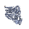
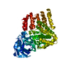
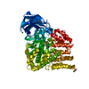
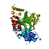
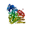
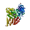
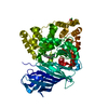
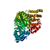
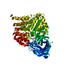
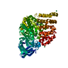
- PDB-3fum: Leukotriene A4 hydrolase in complex with (R)-pyridin-4-yl[4-(2-py... -
+ Open data

- Basic information
| Entry | Database: PDB / ID: 3fum | ||||||
|---|---|---|---|---|---|---|---|
| Title | Leukotriene A4 hydrolase in complex with (R)-pyridin-4-yl[4-(2-pyrrolidin-1-ylethoxy)phenyl]methanol | ||||||
 Components Components | Leukotriene A-4 hydrolase | ||||||
 Keywords Keywords | HYDROLASE / Leukotriene A4 Hydrolase / LTA4H / Fragment crystallography / Fragments of Life / FOL / Alternative splicing / Cytoplasm / Leukotriene biosynthesis / Metal-binding / Metalloprotease / Multifunctional enzyme / Polymorphism / Protease / Zinc | ||||||
| Function / homology |  Function and homology information Function and homology informationleukotriene-A4 hydrolase / leukotriene-A4 hydrolase activity / tripeptide aminopeptidase activity / tripeptide aminopeptidase / Biosynthesis of protectins / Biosynthesis of aspirin-triggered D-series resolvins / Biosynthesis of E-series 18(R)-resolvins / Biosynthesis of D-series resolvins / Biosynthesis of E-series 18(S)-resolvins / Synthesis of Leukotrienes (LT) and Eoxins (EX) ...leukotriene-A4 hydrolase / leukotriene-A4 hydrolase activity / tripeptide aminopeptidase activity / tripeptide aminopeptidase / Biosynthesis of protectins / Biosynthesis of aspirin-triggered D-series resolvins / Biosynthesis of E-series 18(R)-resolvins / Biosynthesis of D-series resolvins / Biosynthesis of E-series 18(S)-resolvins / Synthesis of Leukotrienes (LT) and Eoxins (EX) / epoxide hydrolase activity / leukotriene biosynthetic process / response to zinc ion / peptide catabolic process / metalloaminopeptidase activity / type I pneumocyte differentiation / aminopeptidase activity / lipid metabolic process / response to peptide hormone / tertiary granule lumen / peptidase activity / ficolin-1-rich granule lumen / Neutrophil degranulation / proteolysis / RNA binding / extracellular exosome / extracellular region / zinc ion binding / nucleoplasm / nucleus / cytosol Similarity search - Function | ||||||
| Biological species |  Homo sapiens (human) Homo sapiens (human) | ||||||
| Method |  X-RAY DIFFRACTION / MOLECULAR REPLACEMENT / Resolution: 2.15 Å X-RAY DIFFRACTION / MOLECULAR REPLACEMENT / Resolution: 2.15 Å | ||||||
 Authors Authors | Davies, D.R. | ||||||
 Citation Citation | Journal: J.Med.Chem. / Year: 2009 Title: Discovery of leukotriene A4 hydrolase inhibitors using metabolomics biased fragment crystallography. Authors: Davies, D.R. / Mamat, B. / Magnusson, O.T. / Christensen, J. / Haraldsson, M.H. / Mishra, R. / Pease, B. / Hansen, E. / Singh, J. / Zembower, D. / Kim, H. / Kiselyov, A.S. / Burgin, A.B. / ...Authors: Davies, D.R. / Mamat, B. / Magnusson, O.T. / Christensen, J. / Haraldsson, M.H. / Mishra, R. / Pease, B. / Hansen, E. / Singh, J. / Zembower, D. / Kim, H. / Kiselyov, A.S. / Burgin, A.B. / Gurney, M.E. / Stewart, L.J. | ||||||
| History |
|
- Structure visualization
| Structure viewer | Molecule: MolmilJmol/JSmol |
|---|
- Downloads & links
-Download
| PDBx/mmCIF format | 3fum.cif.gz | 140.2 KB | Display | PDBx/mmCIF format |
|---|---|---|---|---|
| PDB format | pdb3fum.ent.gz | 107 KB | Display | PDB format |
| PDBx/mmJSON format | 3fum.json.gz | Tree view | PDBx/mmJSON format | |
| Others |  Other downloads Other downloads |
-Validation report
| Arichive directory | https://data.pdbj.org/pub/pdb/validation_reports/fu/3fumftp://data.pdbj.org/pub/pdb/validation_reports/fu/3fum | HTTPS FTP |
|---|
-Related structure data
| Related structure data | 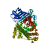 3ftsC 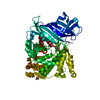 3ftuC 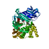 3ftvC 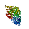 3ftwC 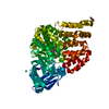 3ftxC  3ftyC 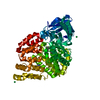 3fu0C  3fu3C 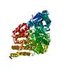 3fu5C  3fu6C  3fudC 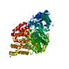 3fueC  3fufC  3fuhC 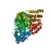 3fuiC 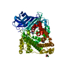 3fujC 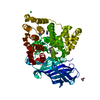 3fukC 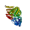 3funC  3fh7S S: Starting model for refinement C: citing same article ( |
|---|---|
| Similar structure data |


-Links
 PDBj
PDBj


- Assembly
| Deposited unit | 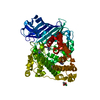
| ||||||||
|---|---|---|---|---|---|---|---|---|---|
| 1 |
| ||||||||
| Unit cell |
|
-Components
-Protein , 1 types, 1 molecules A
| #1: Protein | Mass: 69363.969 Da / Num. of mol.: 1 Source method: isolated from a genetically manipulated source Source: (gene. exp.) Homo sapiens (human) / Gene: LTA4H, LTA4 / Production host:  |
|---|
-Non-polymers , 6 types, 246 molecules 


| #2: Chemical | ChemComp-ZN /  Mass: 65.409 Da / Num. of mol.: 1 / Source method: obtained synthetically / Formula: Zn Mass: 65.409 Da / Num. of mol.: 1 / Source method: obtained synthetically / Formula: Zn | ||
|---|---|---|---|
| #3: Chemical | ChemComp-YB /  Mass: 173.040 Da / Num. of mol.: 1 / Source method: obtained synthetically / Formula: Yb Mass: 173.040 Da / Num. of mol.: 1 / Source method: obtained synthetically / Formula: Yb | ||
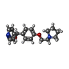
| #4: Chemical | ChemComp-80A / ( Mass: 298.379 Da / Num. of mol.: 1 / Source method: obtained synthetically / Formula: C18H22N2O2 Mass: 298.379 Da / Num. of mol.: 1 / Source method: obtained synthetically / Formula: C18H22N2O2 | ||
| #5: Chemical | ChemComp-ACT /  Mass: 59.044 Da / Num. of mol.: 1 / Source method: obtained synthetically / Formula: C2H3O2 Mass: 59.044 Da / Num. of mol.: 1 / Source method: obtained synthetically / Formula: C2H3O2 | ||
| #6: Chemical |  Mass: 69.085 Da / Num. of mol.: 2 / Source method: obtained synthetically / Formula: C3H5N2 Mass: 69.085 Da / Num. of mol.: 2 / Source method: obtained synthetically / Formula: C3H5N2#7: Water | ChemComp-HOH / | Mass: 18.015 Da / Num. of mol.: 240 / Source method: isolated from a natural source / Formula: H2O |
-Experimental details
-Experiment
| Experiment | Method: X-RAY DIFFRACTION / Number of used crystals: 1 |
|---|
- Sample preparation
| Crystal | Density Matthews: 2.44 Å3/Da / Density % sol: 49.67 % |
|---|---|
| Crystal grow | Temperature: 289 K / Method: vapor diffusion, sitting drop / pH: 6.5 Details: 13% PEG 8000, 100 mM Imidazole pH 6.5, 100 mM Na Acetate, 5 mM YbCl3, crystal soaked with 25 mM (R)-pyridin-4-yl[4-(2-pyrrolidin-1-ylethoxy)phenyl]methanol, VAPOR DIFFUSION, SITTING DROP, temperature 289K |
-Data collection
| Diffraction | Mean temperature: 100 K |
|---|---|
| Diffraction source | Source: ROTATING ANODE / Type: RIGAKU MICROMAX-007 HF / Wavelength: 1.54178 Å |
| Detector | Type: RIGAKU SATURN 944 / Detector: CCD / Date: Sep 26, 2006 |
| Radiation | Protocol: SINGLE WAVELENGTH / Monochromatic (M) / Laue (L): M / Scattering type: x-ray |
| Radiation wavelength | Wavelength: 1.54178 Å / Relative weight: 1 |
| Reflection | Resolution: 2.15→50 Å / Num. obs: 37660 |
- Processing
| Software |
| ||||||||||||||||||||||||||||||||||||||||||||||||||||||||||||||||||||||||||||||||||||||||||||||||||||||||||||||||||||||||||||||||||||||||||||||||||||||||||||||||||||||||||
|---|---|---|---|---|---|---|---|---|---|---|---|---|---|---|---|---|---|---|---|---|---|---|---|---|---|---|---|---|---|---|---|---|---|---|---|---|---|---|---|---|---|---|---|---|---|---|---|---|---|---|---|---|---|---|---|---|---|---|---|---|---|---|---|---|---|---|---|---|---|---|---|---|---|---|---|---|---|---|---|---|---|---|---|---|---|---|---|---|---|---|---|---|---|---|---|---|---|---|---|---|---|---|---|---|---|---|---|---|---|---|---|---|---|---|---|---|---|---|---|---|---|---|---|---|---|---|---|---|---|---|---|---|---|---|---|---|---|---|---|---|---|---|---|---|---|---|---|---|---|---|---|---|---|---|---|---|---|---|---|---|---|---|---|---|---|---|---|---|---|---|---|
| Refinement | Method to determine structure: MOLECULAR REPLACEMENT Starting model: PDB entry 3FH7 Resolution: 2.15→50 Å / Cor.coef. Fo:Fc: 0.948 / Cor.coef. Fo:Fc free: 0.919 / SU B: 5.739 / SU ML: 0.148 / Cross valid method: THROUGHOUT / ESU R: 0.244 / ESU R Free: 0.204 / Stereochemistry target values: MAXIMUM LIKELIHOOD / Details: HYDROGENS HAVE BEEN ADDED IN THE RIDING POSITIONS
| ||||||||||||||||||||||||||||||||||||||||||||||||||||||||||||||||||||||||||||||||||||||||||||||||||||||||||||||||||||||||||||||||||||||||||||||||||||||||||||||||||||||||||
| Solvent computation | Ion probe radii: 0.8 Å / Shrinkage radii: 0.8 Å / VDW probe radii: 1.4 Å / Solvent model: MASK | ||||||||||||||||||||||||||||||||||||||||||||||||||||||||||||||||||||||||||||||||||||||||||||||||||||||||||||||||||||||||||||||||||||||||||||||||||||||||||||||||||||||||||
| Displacement parameters | Biso mean: 24.57 Å2
| ||||||||||||||||||||||||||||||||||||||||||||||||||||||||||||||||||||||||||||||||||||||||||||||||||||||||||||||||||||||||||||||||||||||||||||||||||||||||||||||||||||||||||
| Refinement step | Cycle: LAST / Resolution: 2.15→50 Å
| ||||||||||||||||||||||||||||||||||||||||||||||||||||||||||||||||||||||||||||||||||||||||||||||||||||||||||||||||||||||||||||||||||||||||||||||||||||||||||||||||||||||||||
| Refine LS restraints |
| ||||||||||||||||||||||||||||||||||||||||||||||||||||||||||||||||||||||||||||||||||||||||||||||||||||||||||||||||||||||||||||||||||||||||||||||||||||||||||||||||||||||||||
| LS refinement shell | Resolution: 2.15→2.21 Å / Total num. of bins used: 20
|
