 Movie
Movie Controller

[English] 日本語
 Yorodumi
- PDB-5ncu: Structure of the subtilisin induced serpin-type proteinase inhibi... -
+ Open data

- Basic information
| Entry | Database: PDB / ID: 5ncu | |||||||||||||||||||||
|---|---|---|---|---|---|---|---|---|---|---|---|---|---|---|---|---|---|---|---|---|---|---|
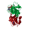
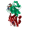
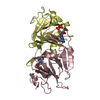
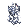
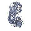
| Title | Structure of the subtilisin induced serpin-type proteinase inhibitor, miropin. | |||||||||||||||||||||
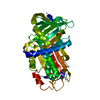
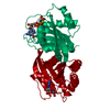
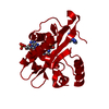
 Components Components | (Serpin) x 2 | |||||||||||||||||||||
 Keywords Keywords | HYDROLASE INHIBITOR / Serpin-type proteinase inhibitor | |||||||||||||||||||||
| Function / homology |  Function and homology information Function and homology information | |||||||||||||||||||||
| Biological species |  Tannerella forsythia (bacteria) Tannerella forsythia (bacteria) | |||||||||||||||||||||
| Method |  X-RAY DIFFRACTION / SYNCHROTRON / MOLECULAR REPLACEMENT / Resolution: 1.7 Å X-RAY DIFFRACTION / SYNCHROTRON / MOLECULAR REPLACEMENT / Resolution: 1.7 Å | |||||||||||||||||||||
 Authors Authors | Goulas, T. / Ksiazek, M. / Garcia-Ferrer, I. / Mizgalska, D. / Potempa, J. / Gomis-Ruth, X. | |||||||||||||||||||||
| Funding support |  Spain, 6items Spain, 6items
| |||||||||||||||||||||
 Citation Citation | Journal: J. Biol. Chem. / Year: 2017 Title: A structure-derived snap-trap mechanism of a multispecific serpin from the dysbiotic human oral microbiome. Authors: Goulas, T. / Ksiazek, M. / Garcia-Ferrer, I. / Sochaj-Gregorczyk, A.M. / Waligorska, I. / Wasylewski, M. / Potempa, J. / Gomis-Ruth, F.X. | |||||||||||||||||||||
| History |
|
- Structure visualization
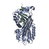
| Structure viewer | Molecule: MolmilJmol/JSmol |
|---|
- Downloads & links
-Download
| PDBx/mmCIF format | 5ncu.cif.gz | 169.3 KB | Display | PDBx/mmCIF format |
|---|---|---|---|---|
| PDB format | pdb5ncu.ent.gz | 131.1 KB | Display | PDB format |
| PDBx/mmJSON format | 5ncu.json.gz | Tree view | PDBx/mmJSON format | |
| Others |  Other downloads Other downloads |
-Validation report
| Arichive directory | https://data.pdbj.org/pub/pdb/validation_reports/nc/5ncuftp://data.pdbj.org/pub/pdb/validation_reports/nc/5ncu | HTTPS FTP |
|---|
-Related structure data
| Related structure data |  5ncsC  5nctC  5ncwC 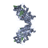 1hleS C: citing same article ( S: Starting model for refinement |
|---|---|
| Similar structure data |

-Links
 PDBj
PDBj
- Assembly
| Deposited unit | 
| ||||||||
|---|---|---|---|---|---|---|---|---|---|
| 1 |
| ||||||||
| Unit cell |
|
-Components
-Protein / Protein/peptide , 2 types, 2 molecules AB
| #1: Protein | Mass: 37705.094 Da / Num. of mol.: 1 Source method: isolated from a genetically manipulated source Details: The amino-terminal residues (GPLGS) are coming from the cloning strategy. Where CSX is S-Oxy cysteine. Source: (gene. exp.) Tannerella forsythia (bacteria) / Gene: BFO_3114 / Production host: |
|---|---|
| #2: Protein/peptide | Mass: 4308.948 Da / Num. of mol.: 1 Source method: isolated from a genetically manipulated source Details: The amino-terminal residues (GPLGS) are coming from the cloning strategy. Where CSX is S-Oxy cysteine. Source: (gene. exp.) Tannerella forsythia (bacteria) / Gene: BFO_3114 / Production host: |
-Non-polymers , 5 types, 414 molecules 


| #3: Chemical | ChemComp-K /  Mass: 39.098 Da / Num. of mol.: 1 / Source method: obtained synthetically / Formula: K Mass: 39.098 Da / Num. of mol.: 1 / Source method: obtained synthetically / Formula: K | ||||||
|---|---|---|---|---|---|---|---|
| #4: Chemical | ChemComp-IOD /  Mass: 126.904 Da / Num. of mol.: 6 / Source method: obtained synthetically / Formula: I Mass: 126.904 Da / Num. of mol.: 6 / Source method: obtained synthetically / Formula: I#5: Chemical | ChemComp-CL / |  Mass: 35.453 Da / Num. of mol.: 1 / Source method: obtained synthetically / Formula: Cl Mass: 35.453 Da / Num. of mol.: 1 / Source method: obtained synthetically / Formula: Cl#6: Chemical | ChemComp-GOL /  Mass: 92.094 Da / Num. of mol.: 4 / Source method: obtained synthetically / Formula: C3H8O3 Mass: 92.094 Da / Num. of mol.: 4 / Source method: obtained synthetically / Formula: C3H8O3#7: Water | ChemComp-HOH / | Mass: 18.015 Da / Num. of mol.: 402 / Source method: isolated from a natural source / Formula: H2O |
-Experimental details
-Experiment
| Experiment | Method: X-RAY DIFFRACTION / Number of used crystals: 1 |
|---|
- Sample preparation
| Crystal | Density Matthews: 2.42 Å3/Da / Density % sol: 49.27 % |
|---|---|
| Crystal grow | Temperature: 293.15 K / Method: vapor diffusion, sitting drop / pH: 7.4 Details: 200 mM sodium iodide 100 mM Bis-Tris, pH 6.5 20% [w/v] polyethylene glycol 3,350 |
-Data collection
| Diffraction | Mean temperature: 100 K |
|---|---|
| Diffraction source | Source: SYNCHROTRON / Site: ALBA / Beamline: XALOC / Wavelength: 0.9793 Å |
| Detector | Type: DECTRIS PILATUS 6M / Detector: PIXEL / Date: Oct 28, 2016 |
| Radiation | Protocol: SINGLE WAVELENGTH / Monochromatic (M) / Laue (L): M / Scattering type: x-ray |
| Radiation wavelength | Wavelength: 0.9793 Å / Relative weight: 1 |
| Reflection | Resolution: 1.7→55.5 Å / Num. obs: 45392 / % possible obs: 99.5 % / Redundancy: 12.1 % / Biso Wilson estimate: 24.51 Å2 / CC1/2: 1 / Rmerge(I) obs: 0.051 / Rrim(I) all: 0.054 / Net I/σ(I): 29.9 |
| Reflection shell | Resolution: 1.7→1.8 Å / Redundancy: 7.6 % / Rmerge(I) obs: 0.479 / Mean I/σ(I) obs: 4.4 / Num. unique all: 3127 / Num. unique obs: 3127 / CC1/2: 0.931 / Rrim(I) all: 0.513 / % possible all: 96.7 |
- Processing
| Software |
| ||||||||||||||||||||||||||||||||||||||||||||||||||||||||||||||||||||||||||||||||||||||||||||||||||||||||||||||||||
|---|---|---|---|---|---|---|---|---|---|---|---|---|---|---|---|---|---|---|---|---|---|---|---|---|---|---|---|---|---|---|---|---|---|---|---|---|---|---|---|---|---|---|---|---|---|---|---|---|---|---|---|---|---|---|---|---|---|---|---|---|---|---|---|---|---|---|---|---|---|---|---|---|---|---|---|---|---|---|---|---|---|---|---|---|---|---|---|---|---|---|---|---|---|---|---|---|---|---|---|---|---|---|---|---|---|---|---|---|---|---|---|---|---|---|---|
| Refinement | Method to determine structure: MOLECULAR REPLACEMENT Starting model: 1HLE Resolution: 1.7→24.89 Å / Cor.coef. Fo:Fc: 0.959 / Cor.coef. Fo:Fc free: 0.948 / Rfactor Rfree error: 0 / SU R Cruickshank DPI: 0.089 / Cross valid method: THROUGHOUT / σ(F): 0 / SU R Blow DPI: 0.097 / SU Rfree Blow DPI: 0.089 / SU Rfree Cruickshank DPI: 0.085
| ||||||||||||||||||||||||||||||||||||||||||||||||||||||||||||||||||||||||||||||||||||||||||||||||||||||||||||||||||
| Displacement parameters | Biso mean: 29.09 Å2
| ||||||||||||||||||||||||||||||||||||||||||||||||||||||||||||||||||||||||||||||||||||||||||||||||||||||||||||||||||
| Refine analyze | Luzzati coordinate error obs: 0.19 Å | ||||||||||||||||||||||||||||||||||||||||||||||||||||||||||||||||||||||||||||||||||||||||||||||||||||||||||||||||||
| Refinement step | Cycle: 1 / Resolution: 1.7→24.89 Å
| ||||||||||||||||||||||||||||||||||||||||||||||||||||||||||||||||||||||||||||||||||||||||||||||||||||||||||||||||||
| Refine LS restraints |
| ||||||||||||||||||||||||||||||||||||||||||||||||||||||||||||||||||||||||||||||||||||||||||||||||||||||||||||||||||
| LS refinement shell | Resolution: 1.7→1.74 Å / Rfactor Rfree error: 0 / Total num. of bins used: 20
| ||||||||||||||||||||||||||||||||||||||||||||||||||||||||||||||||||||||||||||||||||||||||||||||||||||||||||||||||||
| Refinement TLS params. | Method: refined / Refine-ID: X-RAY DIFFRACTION
| ||||||||||||||||||||||||||||||||||||||||||||||||||||||||||||||||||||||||||||||||||||||||||||||||||||||||||||||||||
| Refinement TLS group |
|
