 Movie
Movie Controller


+ Open data

- Basic information
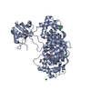
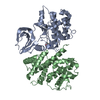
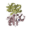
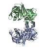
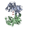
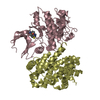
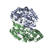
| Entry | Database: PDB / ID: 5n7f | |||||||||
|---|---|---|---|---|---|---|---|---|---|---|
| Title | MAGI-1 complexed with a pRSK1 peptide | |||||||||
 Components Components |
| |||||||||
 Keywords Keywords | SIGNALING PROTEIN / docking / complex | |||||||||
| Function / homology |  Function and homology information Function and homology informationregulation of translation in response to stress / CREB1 phosphorylation through NMDA receptor-mediated activation of RAS signaling / AnxA2-p11 complex / membrane raft assembly / positive regulation of receptor-mediated endocytosis involved in cholesterol transport / positive regulation of vacuole organization / ribosomal protein S6 kinase activity / phospholipase A2 inhibitor activity / positive regulation of low-density lipoprotein particle clearance / hepatocyte proliferation ...regulation of translation in response to stress / CREB1 phosphorylation through NMDA receptor-mediated activation of RAS signaling / AnxA2-p11 complex / membrane raft assembly / positive regulation of receptor-mediated endocytosis involved in cholesterol transport / positive regulation of vacuole organization / ribosomal protein S6 kinase activity / phospholipase A2 inhibitor activity / positive regulation of low-density lipoprotein particle clearance / hepatocyte proliferation / CREB phosphorylation / negative regulation of low-density lipoprotein particle receptor catabolic process / positive regulation of plasma membrane repair / positive regulation of plasminogen activation / positive regulation of hepatic stellate cell activation / PCSK9-AnxA2 complex / myelin sheath adaxonal region / positive regulation of cell-cell adhesion / cadherin binding involved in cell-cell adhesion / positive regulation of vesicle fusion / cornified envelope / endothelial cell morphogenesis / Schmidt-Lanterman incisure / vesicle budding from membrane / Gastrin-CREB signalling pathway via PKC and MAPK / calcium-dependent phospholipid binding / osteoclast development / negative regulation of receptor internalization / plasma membrane protein complex / RSK activation / Dissolution of Fibrin Clot / S100 protein binding / collagen fibril organization / negative regulation of TOR signaling / epithelial cell apoptotic process / vesicle membrane / phosphatidylserine binding / ERK/MAPK targets / Recycling pathway of L1 / positive regulation of receptor recycling / TORC1 signaling / positive regulation of exocytosis / basement membrane / alpha-actinin binding / regulation of neurogenesis / bicellular tight junction / Smooth Muscle Contraction / fibrinolysis / cytoskeletal protein binding / cell projection / phosphatidylinositol-4,5-bisphosphate binding / protein serine/threonine/tyrosine kinase activity / Transcriptional and post-translational regulation of MITF-M expression and activity / lipid droplet / lung development / Gene and protein expression by JAK-STAT signaling after Interleukin-12 stimulation / Turbulent (oscillatory, disturbed) flow shear stress activates signaling by PIEZO1 and integrins in endothelial cells / cell-matrix adhesion / response to activity / Developmental Lineage of Pancreatic Ductal Cells / cell periphery / adherens junction / positive regulation of cell differentiation / serine-type endopeptidase inhibitor activity / sarcolemma / mRNA transcription by RNA polymerase II / RNA polymerase II transcription regulator complex / nuclear matrix / calcium-dependent protein binding / cell-cell junction / azurophil granule lumen / cell junction / melanosome / late endosome membrane / extracellular matrix / positive regulation of cell growth / protease binding / protein-containing complex assembly / Senescence-Associated Secretory Phenotype (SASP) / angiogenesis / midbody / vesicle / basolateral plasma membrane / chemical synaptic transmission / protein phosphorylation / early endosome / non-specific serine/threonine protein kinase / cell surface receptor signaling pathway / cell adhesion / endosome / protein serine kinase activity / lysosomal membrane / protein serine/threonine kinase activity / calcium ion binding / Neutrophil degranulation / regulation of DNA-templated transcription / synapse / negative regulation of apoptotic process / positive regulation of DNA-templated transcription / nucleolus Similarity search - Function | |||||||||
| Biological species |  Homo sapiens (human) Homo sapiens (human) | |||||||||
| Method |  X-RAY DIFFRACTION / SYNCHROTRON / MOLECULAR REPLACEMENT / Resolution: 2.3 Å X-RAY DIFFRACTION / SYNCHROTRON / MOLECULAR REPLACEMENT / Resolution: 2.3 Å | |||||||||
 Authors Authors | Gogl, G. / Nyitray, L. | |||||||||
 Citation Citation | Journal: FEBS J. / Year: 2018 Title: Dynamic control of RSK complexes by phosphoswitch-based regulation. Authors: Gogl, G. / Biri-Kovacs, B. / Poti, A.L. / Vadaszi, H. / Szeder, B. / Bodor, A. / Schlosser, G. / Acs, A. / Turiak, L. / Buday, L. / Alexa, A. / Nyitray, L. / Remenyi, A. | |||||||||
| History |
|
- Structure visualization
| Structure viewer | Molecule: MolmilJmol/JSmol |
|---|
- Downloads & links
-Download
| PDBx/mmCIF format | 5n7f.cif.gz | 365.7 KB | Display | PDBx/mmCIF format |
|---|---|---|---|---|
| PDB format | pdb5n7f.ent.gz | 297.4 KB | Display | PDB format |
| PDBx/mmJSON format | 5n7f.json.gz | Tree view | PDBx/mmJSON format | |
| Others |  Other downloads Other downloads |
-Validation report
| Arichive directory | https://data.pdbj.org/pub/pdb/validation_reports/n7/5n7fftp://data.pdbj.org/pub/pdb/validation_reports/n7/5n7f | HTTPS FTP |
|---|
-Related structure data
| Related structure data |  5n7dC  5n7gC  1xjlS S: Starting model for refinement C: citing same article ( |
|---|---|
| Similar structure data |


-Links
 PDBj
PDBj


- Assembly
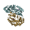
| Deposited unit | 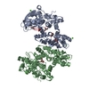
| ||||||||||||
|---|---|---|---|---|---|---|---|---|---|---|---|---|---|
| 1 | 
| ||||||||||||
| 2 | 
| ||||||||||||
| Unit cell |
| ||||||||||||
| Components on special symmetry positions |
|
-Components
| #1: Protein | Mass: 48099.840 Da / Num. of mol.: 2 Source method: isolated from a genetically manipulated source Source: (gene. exp.) Homo sapiens (human)Gene: MAGI1, AIP3, BAIAP1, BAP1, TNRC19, ANXA2, ANX2, ANX2L4, CAL1H, LPC2D Production host:  #2: Protein/peptide | | Mass: 5340.076 Da / Num. of mol.: 1 Source method: isolated from a genetically manipulated source Source: (gene. exp.) Homo sapiens (human) / Gene: RPS6KA1, MAPKAPK1A, RSK1 / Production host: References: UniProt: Q15418, non-specific serine/threonine protein kinase #3: Chemical | ChemComp-GOL /   Mass: 92.094 Da / Num. of mol.: 6 / Source method: obtained synthetically / Formula: C3H8O3 Mass: 92.094 Da / Num. of mol.: 6 / Source method: obtained synthetically / Formula: C3H8O3#4: Chemical | ChemComp-CA /   Mass: 40.078 Da / Num. of mol.: 9 / Source method: obtained synthetically / Formula: Ca Mass: 40.078 Da / Num. of mol.: 9 / Source method: obtained synthetically / Formula: Ca#5: Water | ChemComp-HOH / |  Mass: 18.015 Da / Num. of mol.: 634 / Source method: isolated from a natural source / Formula: H2O Mass: 18.015 Da / Num. of mol.: 634 / Source method: isolated from a natural source / Formula: H2OHas protein modification | Y | |
|---|
-Experimental details
-Experiment
| Experiment | Method: X-RAY DIFFRACTION / Number of used crystals: 1 |
|---|
- Sample preparation
| Crystal | Density Matthews: 2.92 Å3/Da / Density % sol: 57.93 % |
|---|---|
| Crystal grow | Temperature: 296 K / Method: vapor diffusion, sitting drop / pH: 8.5 / Details: 14% PEG 8000, 200 mM MgCl2, 100 mM TRIS |
-Data collection
| Diffraction | Mean temperature: 100 K |
|---|---|
| Diffraction source | Source: SYNCHROTRON / Site: SLS  / Beamline: X06DA / Wavelength: 1 Å / Beamline: X06DA / Wavelength: 1 Å |
| Detector | Type: DECTRIS PILATUS 2M / Detector: PIXEL / Date: Dec 3, 2016 |
| Radiation | Protocol: SINGLE WAVELENGTH / Monochromatic (M) / Laue (L): M / Scattering type: x-ray |
| Radiation wavelength | Wavelength: 1 Å / Relative weight: 1 |
| Reflection | Resolution: 2.3→49.73 Å / Num. obs: 53922 / % possible obs: 100 % / Redundancy: 19.87 % / Rrim(I) all: 0.219 / Net I/σ(I): 15.42 |
| Reflection shell | Resolution: 2.3→2.36 Å / Redundancy: 18.6 % / Mean I/σ(I) obs: 3 / Num. unique all: 3925 / % possible all: 100 |
- Processing
| Software |
| ||||||||||||||||||||||||||||||||||||||||||||||||||||||||||||||||||||||||||||||||||||||||||||||||||||||||||||||||||||||||||||||||||||||||||||||||||||||
|---|---|---|---|---|---|---|---|---|---|---|---|---|---|---|---|---|---|---|---|---|---|---|---|---|---|---|---|---|---|---|---|---|---|---|---|---|---|---|---|---|---|---|---|---|---|---|---|---|---|---|---|---|---|---|---|---|---|---|---|---|---|---|---|---|---|---|---|---|---|---|---|---|---|---|---|---|---|---|---|---|---|---|---|---|---|---|---|---|---|---|---|---|---|---|---|---|---|---|---|---|---|---|---|---|---|---|---|---|---|---|---|---|---|---|---|---|---|---|---|---|---|---|---|---|---|---|---|---|---|---|---|---|---|---|---|---|---|---|---|---|---|---|---|---|---|---|---|---|---|---|---|
| Refinement | Method to determine structure: MOLECULAR REPLACEMENT Starting model: 1xjl Resolution: 2.3→49.73 Å / SU ML: 0.31 / Cross valid method: FREE R-VALUE / σ(F): 1.34 / Phase error: 23.27
| ||||||||||||||||||||||||||||||||||||||||||||||||||||||||||||||||||||||||||||||||||||||||||||||||||||||||||||||||||||||||||||||||||||||||||||||||||||||
| Solvent computation | Shrinkage radii: 0.9 Å / VDW probe radii: 1.11 Å | ||||||||||||||||||||||||||||||||||||||||||||||||||||||||||||||||||||||||||||||||||||||||||||||||||||||||||||||||||||||||||||||||||||||||||||||||||||||
| Refinement step | Cycle: LAST / Resolution: 2.3→49.73 Å
| ||||||||||||||||||||||||||||||||||||||||||||||||||||||||||||||||||||||||||||||||||||||||||||||||||||||||||||||||||||||||||||||||||||||||||||||||||||||
| Refine LS restraints |
| ||||||||||||||||||||||||||||||||||||||||||||||||||||||||||||||||||||||||||||||||||||||||||||||||||||||||||||||||||||||||||||||||||||||||||||||||||||||
| LS refinement shell |
| ||||||||||||||||||||||||||||||||||||||||||||||||||||||||||||||||||||||||||||||||||||||||||||||||||||||||||||||||||||||||||||||||||||||||||||||||||||||
| Refinement TLS params. | Method: refined / Refine-ID: X-RAY DIFFRACTION
| ||||||||||||||||||||||||||||||||||||||||||||||||||||||||||||||||||||||||||||||||||||||||||||||||||||||||||||||||||||||||||||||||||||||||||||||||||||||
| Refinement TLS group |
|
