 Movie
Movie Controller

[English] 日本語
 Yorodumi
- PDB-5n1t: Crystal structure of complex between flavocytochrome c and copper... -
+ Open data

- Basic information
| Entry | Database: PDB / ID: 5n1t | ||||||
|---|---|---|---|---|---|---|---|
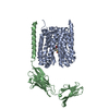
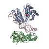
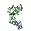
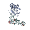
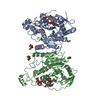
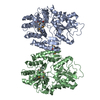
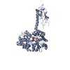
| Title | Crystal structure of complex between flavocytochrome c and copper chaperone CopC from T. paradoxus | ||||||
 Components Components |
| ||||||
 Keywords Keywords | OXIDOREDUCTASE / COPPER CHAPERONE / SULFIDE REDUCTASE / PERIPLASM / PROTEIN COMPLEX | ||||||
| Function / homology |  Function and homology information Function and homology informationresponse to copper ion / flavin adenine dinucleotide binding / periplasmic space / electron transfer activity / oxidoreductase activity / copper ion binding / heme binding / metal ion binding Similarity search - Function | ||||||
| Biological species |  Thioalkalivibrio paradoxus ARh 1 (bacteria) Thioalkalivibrio paradoxus ARh 1 (bacteria) | ||||||
| Method |  X-RAY DIFFRACTION / SYNCHROTRON / MOLECULAR REPLACEMENT / Resolution: 2.6 Å X-RAY DIFFRACTION / SYNCHROTRON / MOLECULAR REPLACEMENT / Resolution: 2.6 Å | ||||||
 Authors Authors | Osipov, E.M. / Lilina, A.V. / Tikhonova, T.V. / Tsallagov, S.I. / Popov, V.O. | ||||||
| Funding support |  Russian Federation, 1items Russian Federation, 1items
| ||||||
 Citation Citation | Journal: Acta Crystallogr D Struct Biol / Year: 2018 Title: Structure of the flavocytochrome c sulfide dehydrogenase associated with the copper-binding protein CopC from the haloalkaliphilic sulfur-oxidizing bacterium Thioalkalivibrio paradoxusARh 1. Authors: Osipov, E.M. / Lilina, A.V. / Tsallagov, S.I. / Safonova, T.N. / Sorokin, D.Y. / Tikhonova, T.V. / Popov, V.O. | ||||||
| History |
|
- Structure visualization
| Structure viewer | Molecule: MolmilJmol/JSmol |
|---|
- Downloads & links
-Download
| PDBx/mmCIF format | 5n1t.cif.gz | 158.4 KB | Display | PDBx/mmCIF format |
|---|---|---|---|---|
| PDB format | pdb5n1t.ent.gz | 117.2 KB | Display | PDB format |
| PDBx/mmJSON format | 5n1t.json.gz | Tree view | PDBx/mmJSON format | |
| Others |  Other downloads Other downloads |
-Validation report
| Arichive directory | https://data.pdbj.org/pub/pdb/validation_reports/n1/5n1tftp://data.pdbj.org/pub/pdb/validation_reports/n1/5n1t | HTTPS FTP |
|---|
-Related structure data
| Related structure data |  1fcdS S: Starting model for refinement |
|---|---|
| Similar structure data |

-Links
 PDBj
PDBj


- Assembly
| Deposited unit | 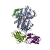
| ||||||||
|---|---|---|---|---|---|---|---|---|---|
| 1 |
| ||||||||
| Unit cell |
|
-Components
-Protein , 3 types, 4 molecules ABMW
| #1: Protein | Mass: 46264.730 Da / Num. of mol.: 1 / Mutation: C199CSS / Source method: isolated from a natural source Source: (natural) Thioalkalivibrio paradoxus ARh 1 (bacteria)References: UniProt: W0DP88 |
|---|---|
| #2: Protein | Mass: 10628.845 Da / Num. of mol.: 1 / Source method: isolated from a natural source Source: (natural) Thioalkalivibrio paradoxus ARh 1 (bacteria)References: UniProt: W0DKT4 |
| #3: Protein | Mass: 16935.814 Da / Num. of mol.: 2 / Source method: isolated from a natural source Source: (natural) Thioalkalivibrio paradoxus ARh 1 (bacteria)References: UniProt: W0DSL1 |
-Non-polymers , 5 types, 61 molecules 


| #4: Chemical | ChemComp-FAD /  Mass: 785.550 Da / Num. of mol.: 1 / Source method: obtained synthetically / Formula: C27H33N9O15P2 / Comment: FAD*YM Mass: 785.550 Da / Num. of mol.: 1 / Source method: obtained synthetically / Formula: C27H33N9O15P2 / Comment: FAD*YM |
|---|---|
| #5: Chemical | ChemComp-HEC /  Mass: 618.503 Da / Num. of mol.: 1 / Source method: obtained synthetically / Formula: C34H34FeN4O4 Mass: 618.503 Da / Num. of mol.: 1 / Source method: obtained synthetically / Formula: C34H34FeN4O4 |
| #6: Chemical | ChemComp-CU /  Mass: 63.546 Da / Num. of mol.: 1 / Source method: obtained synthetically / Formula: Cu Mass: 63.546 Da / Num. of mol.: 1 / Source method: obtained synthetically / Formula: Cu |
| #7: Chemical | ChemComp-GOL /  Mass: 92.094 Da / Num. of mol.: 1 / Source method: obtained synthetically / Formula: C3H8O3 Mass: 92.094 Da / Num. of mol.: 1 / Source method: obtained synthetically / Formula: C3H8O3 |
| #8: Water | ChemComp-HOH / Mass: 18.015 Da / Num. of mol.: 57 / Source method: isolated from a natural source / Formula: H2O |
-Details
| Has protein modification | Y |
|---|
-Experimental details
-Experiment
| Experiment | Method: X-RAY DIFFRACTION / Number of used crystals: 1 |
|---|
- Sample preparation
| Crystal | Density Matthews: 2.35 Å3/Da / Density % sol: 47.61 % |
|---|---|
| Crystal grow | Temperature: 293 K / Method: vapor diffusion, hanging drop / pH: 8.2 Details: 0.056 M Sodium phosphate monobasic monohydrate, 1.344 M Potassium phosphate dibasic |
-Data collection
| Diffraction | Mean temperature: 100 K |
|---|---|
| Diffraction source | Source: SYNCHROTRON / Site: ESRF  / Beamline: ID23-1 / Wavelength: 0.976256 Å / Beamline: ID23-1 / Wavelength: 0.976256 Å |
| Detector | Type: DECTRIS PILATUS3 S 6M / Detector: PIXEL / Date: Feb 14, 2015 |
| Radiation | Protocol: SINGLE WAVELENGTH / Monochromatic (M) / Laue (L): M / Scattering type: x-ray |
| Radiation wavelength | Wavelength: 0.976256 Å / Relative weight: 1 |
| Reflection | Resolution: 2.6→30 Å / Num. obs: 28526 / % possible obs: 93.1 % / Redundancy: 3.7 % / Biso Wilson estimate: 56 Å2 / CC1/2: 0.99 / Rrim(I) all: 0.117 / Net I/σ(I): 9.8 |
| Reflection shell | Resolution: 2.6→2.7 Å / Redundancy: 5.5 % / Mean I/σ(I) obs: 2.1 / Num. unique obs: 2841 / CC1/2: 0.79 / Rrim(I) all: 0.91 / % possible all: 99.8 |
- Processing
| Software |
| ||||||||||||||||||||||||||||||||||||||||||||||||||||||||||||||||||||||||||||||||||||||||||||||||||||||||||||||||||||||||||||||||||||||||||||||||||||||||||||||||||||||||||||||||||||||
|---|---|---|---|---|---|---|---|---|---|---|---|---|---|---|---|---|---|---|---|---|---|---|---|---|---|---|---|---|---|---|---|---|---|---|---|---|---|---|---|---|---|---|---|---|---|---|---|---|---|---|---|---|---|---|---|---|---|---|---|---|---|---|---|---|---|---|---|---|---|---|---|---|---|---|---|---|---|---|---|---|---|---|---|---|---|---|---|---|---|---|---|---|---|---|---|---|---|---|---|---|---|---|---|---|---|---|---|---|---|---|---|---|---|---|---|---|---|---|---|---|---|---|---|---|---|---|---|---|---|---|---|---|---|---|---|---|---|---|---|---|---|---|---|---|---|---|---|---|---|---|---|---|---|---|---|---|---|---|---|---|---|---|---|---|---|---|---|---|---|---|---|---|---|---|---|---|---|---|---|---|---|---|---|
| Refinement | Method to determine structure: MOLECULAR REPLACEMENT Starting model: 1FCD Resolution: 2.6→30 Å / Cor.coef. Fo:Fc: 0.959 / Cor.coef. Fo:Fc free: 0.921 / SU B: 12.985 / SU ML: 0.261 / Cross valid method: THROUGHOUT / ESU R: 0.58 / ESU R Free: 0.313 / Stereochemistry target values: MAXIMUM LIKELIHOOD
| ||||||||||||||||||||||||||||||||||||||||||||||||||||||||||||||||||||||||||||||||||||||||||||||||||||||||||||||||||||||||||||||||||||||||||||||||||||||||||||||||||||||||||||||||||||||
| Solvent computation | Ion probe radii: 0.8 Å / Shrinkage radii: 0.8 Å / VDW probe radii: 1.2 Å / Solvent model: BABINET MODEL WITH MASK | ||||||||||||||||||||||||||||||||||||||||||||||||||||||||||||||||||||||||||||||||||||||||||||||||||||||||||||||||||||||||||||||||||||||||||||||||||||||||||||||||||||||||||||||||||||||
| Displacement parameters | Biso mean: 55.429 Å2
| ||||||||||||||||||||||||||||||||||||||||||||||||||||||||||||||||||||||||||||||||||||||||||||||||||||||||||||||||||||||||||||||||||||||||||||||||||||||||||||||||||||||||||||||||||||||
| Refinement step | Cycle: 1 / Resolution: 2.6→30 Å
| ||||||||||||||||||||||||||||||||||||||||||||||||||||||||||||||||||||||||||||||||||||||||||||||||||||||||||||||||||||||||||||||||||||||||||||||||||||||||||||||||||||||||||||||||||||||
| Refine LS restraints |
|