[English] 日本語
Yorodumi
- PDB-5n06: Crystal structure of Tie1 Fibronectin-like domain 3 -

+
Open data


ID or keywords:

Loading...

-
Basic information

Entry
Database: PDB / ID: 5n06
TitleCrystal structure of Tie1 Fibronectin-like domain 3
ComponentsTyrosine-protein kinase receptor Tie-1
KeywordsSIGNALING PROTEIN / receptor / fibronectin-like domains / heterodimerization
Function / homology
Function and homology information


branching involved in lymph vessel morphogenesis / plasma membrane fusion / lymphatic endothelial cell differentiation / regulation of endothelial cell proliferation / regulation of extracellular matrix assembly / tissue remodeling / multicellular organism development / positive regulation of kinase activity / aortic valve morphogenesis / plasma membrane => GO:0005886 ...branching involved in lymph vessel morphogenesis / plasma membrane fusion / lymphatic endothelial cell differentiation / regulation of endothelial cell proliferation / regulation of extracellular matrix assembly / tissue remodeling / multicellular organism development / positive regulation of kinase activity / aortic valve morphogenesis / plasma membrane => GO:0005886 / mesoderm development / vasculogenesis / response to retinoic acid / cell surface receptor protein tyrosine kinase signaling pathway / transmembrane receptor protein tyrosine kinase activity / negative regulation of angiogenesis / negative regulation of cell migration / receptor protein-tyrosine kinase / positive regulation of angiogenesis / angiogenesis / in utero embryonic development / receptor complex / signal transduction / ATP binding
Similarity search - Function
Immunoglobulin / Immunoglobulin domain / Epidermal growth factor-like domain. / EGF-like domain profile. / EGF-like domain signature 2. / Fibronectin type III domain / EGF-like domain signature 1. / EGF-like domain / Fibronectin type 3 domain / Fibronectin type-III domain profile. ...Immunoglobulin / Immunoglobulin domain / Epidermal growth factor-like domain. / EGF-like domain profile. / EGF-like domain signature 2. / Fibronectin type III domain / EGF-like domain signature 1. / EGF-like domain / Fibronectin type 3 domain / Fibronectin type-III domain profile. / Fibronectin type III / Fibronectin type III superfamily / Tyrosine-protein kinase, catalytic domain / Tyrosine kinase, catalytic domain / Tyrosine protein kinases specific active-site signature. / Immunoglobulin subtype / Immunoglobulin / Tyrosine-protein kinase, active site / Protein tyrosine and serine/threonine kinase / Serine-threonine/tyrosine-protein kinase, catalytic domain / Immunoglobulin-like domain superfamily / Protein kinase, ATP binding site / Protein kinases ATP-binding region signature. / Immunoglobulin-like fold / Protein kinase domain profile. / Protein kinase domain / Protein kinase-like domain superfamily
Similarity search - Domain/homology
Tyrosine-protein kinase receptor Tie-1
Similarity search - Component
Biological speciesHomo sapiens (human)
MethodX-RAY DIFFRACTION / SYNCHROTRON / MOLECULAR REPLACEMENT / Resolution: 2.501 Å
AuthorsLeppanen, V.-M. / Saharinen, P. / Alitalo, K.
CitationJournal: Proc. Natl. Acad. Sci. U.S.A. / Year: 2017
Title: Structural basis of Tie2 activation and Tie2/Tie1 heterodimerization.
Authors: Leppanen, V.M. / Saharinen, P. / Alitalo, K.
History
DepositionFeb 2, 2017Deposition site: PDBE / Processing site: PDBE
Revision 1.0Apr 26, 2017Provider: repository / Type: Initial release
Revision 1.1May 3, 2017Group: Database references
Revision 1.2May 1, 2024Group: Data collection / Database references / Refinement description
Category: chem_comp_atom / chem_comp_bond ...chem_comp_atom / chem_comp_bond / database_2 / pdbx_initial_refinement_model
Item: _database_2.pdbx_DOI / _database_2.pdbx_database_accession

-
Structure visualization

Structure viewerMolecule:
MolmilJmol/JSmol

Downloads & links

-
Assembly

Deposited unit
A: Tyrosine-protein kinase receptor Tie-1
B: Tyrosine-protein kinase receptor Tie-1


Theoretical massNumber of molelcules
Total (without water)28,1342
Polymers28,1342
Non-polymers00
Water1,31573
1


  • Idetical with deposited unit
  • defined by software
TypeNameSymmetry operationNumber
identity operation1_555x,y,z1
Buried area3930 Å2
ΔGint-16 kcal/mol
Surface area10010 Å2
MethodPISA
Unit cell
Length a, b, c (Å)54.054, 54.054, 107.270
Angle α, β, γ (deg.)90.00, 90.00, 120.00
Int Tables number170
Space group name H-MP65

-
Components

#1: Protein Tyrosine-protein kinase receptor Tie-1


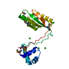
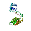
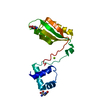
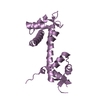
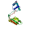
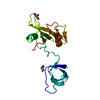
Mass: 14066.904 Da / Num. of mol.: 2
Source method: isolated from a genetically manipulated source
Details: The sequence includes N-terminal Mellitin signal peptide, cloning artefect (ADP) and C-terminal His-tag.
Source: (gene. exp.) Homo sapiens (human) / Gene: TIE1, TIE
Production host: 
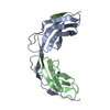
Spodoptera aff. frugiperda 2 RZ-2014 (butterflies/moths)
References: UniProt: P35590, receptor protein-tyrosine kinase
#2: Water ChemComp-HOH / water


Mass: 18.015 Da / Num. of mol.: 73 / Source method: isolated from a natural source / Formula: H2O

-
Experimental details

-
Experiment

ExperimentMethod: X-RAY DIFFRACTION / Number of used crystals: 1

-
Sample preparation

CrystalDensity Matthews: 2 Å3/Da / Density % sol: 37 % / Description: Hexagonal rods
Crystal growTemperature: 294 K / Method: vapor diffusion, sitting drop
Details: 0.1 M Tris buffer at pH 8.0 - 9.0 and 18-24% PEG 8000 (w/v)
PH range: 8.0 - 9.0 / Temp details: Room temperature

-
Data collection

DiffractionMean temperature: 100 K / Ambient temp details: Liguid nitrogen cooling system
Diffraction sourceSource: SYNCHROTRON / Site: ESRF / Beamline: ID29 / Wavelength: 0.97895 Å
DetectorType: DECTRIS PILATUS 6M / Detector: PIXEL / Date: Jul 2, 2014
RadiationMonochromator: liquid nitrogen cooled channel-cut silicon monochromator
Protocol: SINGLE WAVELENGTH / Monochromatic (M) / Laue (L): M / Scattering type: x-ray
Radiation wavelengthWavelength: 0.97895 Å / Relative weight: 1
ReflectionResolution: 2.5→30 Å / Num. obs: 6161 / % possible obs: 99.6 % / Redundancy: 7.6 % / Biso Wilson estimate: 79.9 Å2 / Rsym value: 0.078 / Net I/σ(I): 15.2
Reflection shellResolution: 2.5→2.65 Å / Redundancy: 6.5 % / Mean I/σ(I) obs: 2 / Num. unique obs: 951 / Rsym value: 0.935 / % possible all: 97.7

-
Processing

Software
NameVersionClassification
PHENIX1.9_1692refinement
XDSdata reduction
XSCALEdata scaling
PHENIXphasing
RefinementMethod to determine structure: MOLECULAR REPLACEMENT
Starting model: Tie2 Fn3 domain

Resolution: 2.501→28.416 Å / Cross valid method: FREE R-VALUE / σ(F): 1.41 / Phase error: 33.97
Details: Maximum likelihood refinement of XYZ coordinates, TLS parameters and individual B-factors. Twin law: k, h, -l. Twin fraction 0.510.
RfactorNum. reflection% reflectionSelection details
Rfree0.3151 370 6.01 %Random selection
Rwork0.2435 ---
obs0.2489 6161 99.97 %-
Solvent computationShrinkage radii: 0.9 Å / VDW probe radii: 1.11 Å
Displacement parametersBiso mean: 107.6 Å2
Refinement stepCycle: LAST / Resolution: 2.501→28.416 Å
ProteinNucleic acidLigandSolventTotal
Num. atoms1339 0 0 73 1412
Refine LS restraints
Refine-IDTypeDev idealNumber
X-RAY DIFFRACTIONf_bond_d0.0051374
X-RAY DIFFRACTIONf_angle_d0.7871889
X-RAY DIFFRACTIONf_dihedral_angle_d11.656471
X-RAY DIFFRACTIONf_chiral_restr0.028221
X-RAY DIFFRACTIONf_plane_restr0.004243
LS refinement shell
Resolution (Å)Rfactor RfreeNum. reflection RfreeRfactor RworkNum. reflection RworkRefine-ID% reflection obs (%)
2.5022-2.8640.32691230.32111924X-RAY DIFFRACTION94
2.864-3.60720.34021230.28261918X-RAY DIFFRACTION94
3.6072-28.41740.30621240.21891939X-RAY DIFFRACTION94
Refinement TLS params.

Method: refined / Refine-ID: X-RAY DIFFRACTION

IDL112)L122)L132)L222)L232)L332)S11 (Å °)S12 (Å °)S13 (Å °)S21 (Å °)S22 (Å °)S23 (Å °)S31 (Å °)S32 (Å °)S33 (Å °)T112)T122)T132)T222)T232)T332)Origin x (Å)Origin y (Å)Origin z (Å)
12.0925-0.33064.95367.74970.58652.0736-1.2161-0.0317-1.7636-0.07240.99910.09321.29132.8104-0.01970.92310.31130.11871.6295-0.1361.09148.2116-19.0932.543
24.9125-1.7980.81113.17810.67743.37360.77071.2248-1.91760.82070.65781.3730.2194-0.3429-0.64380.8911.40731.06882.130.61881.6832-6.5935-18.2148-5.7205
32.07241.4377.0832.0434-2.7372.01431.9042-0.5914-0.9720.6187-0.41540.4520.3491-0.7413-1.51570.6455-0.00660.05331.0919-0.03420.57510.0604-12.78044.0302
44.88491.3963-2.5693.84784.69349.83521.1129-1.6351-0.76590.26421.4578-1.0382-0.3807-1.8986-2.33750.66710.0214-0.00120.98440.21351.03272.8949-10.339911.0151
54.7469-1.9371-3.19017.25712.06442.0155-0.2390.09210.6035-0.38361.6507-1.1351-0.827-0.2518-1.46140.625-0.2020.04050.999300.73134.3904-13.0099-4.391
62.2893-5.19045.62265.2362-2.5967.0201-1.7618-0.46320.76061.54361.1314-0.2964-1.39120.5410.6230.91720.1561-0.06721.00980.04610.64252.4028-12.95776.3958
70.7175-1.82011.15382.04240.65363.8070.0049-0.51990.119-0.39060.01071.8418-1.46840.5862-0.17771.30220.13660.02190.94810.11141.1695-14.999515.3818-9.24
83.7343-1.59130.63088.07165.5738.05870.19810.42890.7168-0.7536-0.43681.2125-1.983-0.10980.26991.01410.1192-0.22870.70440.00891.0415-17.375313.2731-2.0007
92.01936.53254.01612.1165-6.30337.88592.79942.05411.4762-2.4577-0.2952-0.7293-0.89423.0441-1.8281.46-0.03470.62171.373-0.22041.1551-5.861820.193-4.0448
109.32081.466-2.89226.2972-2.82824.7671.42110.63280.23220.7259-0.57882.3172-1.4747-1.274-0.54740.92970.20310.2060.8309-0.02230.8277-18.98253.45334.7733
112.0211-1.23821.9042.0321-6.62732.02480.0271.62111.3974-0.84390.34872.4698-1.27750.2569-0.05551.2181-0.1167-0.07720.96580.3011.0857-10.8496.6669-6.4084
124.10420.2599-0.15783.13550.25720.87550.2591.31780.69950.60820.51970.0352-0.3049-0.4081-0.76671.28640.77710.3511.74180.2941.0114-6.83526.438-8.9623
134.80750.2698-1.28327.0422-0.63762.71011.5438-1.62371.02781.32-1.0343-0.293-0.23290.8166-0.42811.6938-0.2833-0.13790.9415-0.30661.0356-8.51429.78863.4782
141.9559-3.6632-1.06151.74850.8530.51071.34722.21412.4114-0.60652.13371.435-2.43360.0483-2.65373.1027-1.4744-0.38862.78610.52340.9735-4.690219.0911-13.1438
153.7329-0.33890.82822.93590.87230.5172-1.30591.34540.9099-0.5544-0.1807-0.5494-0.6977-0.55331.21062.34550.2299-0.0080.7479-0.15310.463-14.54987.6024-6.924
163.2218-0.43720.38365.6641.32380.44231.1213-0.829-1.4575-0.0414-1.53780.02950.42831.04670.15710.79030.3272-0.01952.02190.42760.92740.2692-17.67627.0749
Refinement TLS group
IDRefine-IDRefine TLS-IDSelection details
1X-RAY DIFFRACTION1chain 'A' and (resid 644 through 663 )
2X-RAY DIFFRACTION2chain 'A' and (resid 664 through 675 )
3X-RAY DIFFRACTION3chain 'A' and (resid 676 through 681 )
4X-RAY DIFFRACTION4chain 'A' and (resid 682 through 695 )
5X-RAY DIFFRACTION5chain 'A' and (resid 696 through 705 )
6X-RAY DIFFRACTION6chain 'A' and (resid 706 through 726 )
7X-RAY DIFFRACTION7chain 'A' and (resid 727 through 738 )
8X-RAY DIFFRACTION8chain 'B' and (resid 644 through 655 )
9X-RAY DIFFRACTION9chain 'B' and (resid 656 through 663 )
10X-RAY DIFFRACTION10chain 'B' and (resid 664 through 675 )
11X-RAY DIFFRACTION11chain 'B' and (resid 676 through 682 )
12X-RAY DIFFRACTION12chain 'B' and (resid 683 through 695 )
13X-RAY DIFFRACTION13chain 'B' and (resid 696 through 705 )
14X-RAY DIFFRACTION14chain 'B' and (resid 706 through 714 )
15X-RAY DIFFRACTION15chain 'B' and (resid 715 through 721 )
16X-RAY DIFFRACTION16chain 'B' and (resid 722 through 737 )

+
About Yorodumi

-
News

-
Feb 9, 2022. New format data for meta-information of EMDB entries

New format data for meta-information of EMDB entries

  • Version 3 of the EMDB header file is now the official format.
  • The previous official version 1.9 will be removed from the archive.

Related info.:EMDB header

External links:wwPDB to switch to version 3 of the EMDB data model

-
Aug 12, 2020. Covid-19 info

Covid-19 info

URL: https://pdbjlvh1.pdbj.org/emnavi/covid19.php

New page: Covid-19 featured information page in EM Navigator.

Related info.:Covid-19 info / Mar 5, 2020. Novel coronavirus structure data

+
Mar 5, 2020. Novel coronavirus structure data

Novel coronavirus structure data

Related info.:Yorodumi Speices / Aug 12, 2020. Covid-19 info

External links:COVID-19 featured content - PDBj / Molecule of the Month (242):Coronavirus Proteases

+
Jan 31, 2019. EMDB accession codes are about to change! (news from PDBe EMDB page)

EMDB accession codes are about to change! (news from PDBe EMDB page)

  • The allocation of 4 digits for EMDB accession codes will soon come to an end. Whilst these codes will remain in use, new EMDB accession codes will include an additional digit and will expand incrementally as the available range of codes is exhausted. The current 4-digit format prefixed with “EMD-” (i.e. EMD-XXXX) will advance to a 5-digit format (i.e. EMD-XXXXX), and so on. It is currently estimated that the 4-digit codes will be depleted around Spring 2019, at which point the 5-digit format will come into force.
  • The EM Navigator/Yorodumi systems omit the EMD- prefix.

Related info.:Q: What is EMD? / ID/Accession-code notation in Yorodumi/EM Navigator

External links:EMDB Accession Codes are Changing Soon! / Contact to PDBj

+
Jul 12, 2017. Major update of PDB

Major update of PDB

  • wwPDB released updated PDB data conforming to the new PDBx/mmCIF dictionary.
  • This is a major update changing the version number from 4 to 5, and with Remediation, in which all the entries are updated.
  • In this update, many items about electron microscopy experimental information are reorganized (e.g. em_software).
  • Now, EM Navigator and Yorodumi are based on the updated data.

External links:wwPDB Remediation / Enriched Model Files Conforming to OneDep Data Standards Now Available in the PDB FTP Archive

-
Yorodumi

Thousand views of thousand structures

  • Yorodumi is a browser for structure data from EMDB, PDB, SASBDB, etc.
  • This page is also the successor to EM Navigator detail page, and also detail information page/front-end page for Omokage search.
  • The word "yorodu" (or yorozu) is an old Japanese word meaning "ten thousand". "mi" (miru) is to see.

Related info.:EMDB / PDB / SASBDB / Comparison of 3 databanks / Yorodumi Search / Aug 31, 2016. New EM Navigator & Yorodumi / Yorodumi Papers / Jmol/JSmol / Function and homology information / Changes in new EM Navigator and Yorodumi

Read more