[English] 日本語
Yorodumi
- PDB-4d8k: Crystal structure of a SH3-SH2 domains of a lymphocyte-specific p... -

+
Open data


ID or keywords:

Loading...

-
Basic information

Entry
Database: PDB / ID: 4d8k
TitleCrystal structure of a SH3-SH2 domains of a lymphocyte-specific protein tyrosine kinase (LCK) from Homo sapiens at 2.36 A resolution
ComponentsTyrosine-protein kinase Lck
KeywordsTRANSFERASE / Protein kinases / SH2 domain / SH3 domain / Structural Genomics / Joint Center for Structural Genomics / JCSG / Protein Structure Initiative / PSI-BIOLOGY / Partnership for T-Cell Biology / TCELL
Function / homology
Function and homology information


regulation of lymphocyte activation / positive regulation of leukocyte cell-cell adhesion / Fc-gamma receptor signaling pathway / CD28 co-stimulation / intracellular zinc ion homeostasis / FLT3 signaling through SRC family kinases / CD4 receptor binding / Nef Mediated CD4 Down-regulation / Nef and signal transduction / positive regulation of heterotypic cell-cell adhesion ...regulation of lymphocyte activation / positive regulation of leukocyte cell-cell adhesion / Fc-gamma receptor signaling pathway / CD28 co-stimulation / intracellular zinc ion homeostasis / FLT3 signaling through SRC family kinases / CD4 receptor binding / Nef Mediated CD4 Down-regulation / Nef and signal transduction / positive regulation of heterotypic cell-cell adhesion / Interleukin-2 signaling / CD28 dependent Vav1 pathway / Regulation of KIT signaling / CTLA4 inhibitory signaling / protein serine/threonine phosphatase activity / leukocyte migration / phospholipase activator activity / CD8 receptor binding / pericentriolar material / positive regulation of T cell receptor signaling pathway / Translocation of ZAP-70 to Immunological synapse / Phosphorylation of CD3 and TCR zeta chains / PECAM1 interactions / CD28 dependent PI3K/Akt signaling / phospholipase binding / RHOH GTPase cycle / hemopoiesis / Generation of second messenger molecules / immunological synapse / T cell differentiation / PD-1 signaling / phosphatidylinositol 3-kinase binding / T cell receptor binding / positive regulation of intrinsic apoptotic signaling pathway / peptidyl-tyrosine autophosphorylation / release of sequestered calcium ion into cytosol / extrinsic component of cytoplasmic side of plasma membrane / GPVI-mediated activation cascade / cell surface receptor protein tyrosine kinase signaling pathway / T cell costimulation / phosphotyrosine residue binding / SH2 domain binding / Signaling by phosphorylated juxtamembrane, extracellular and kinase domain KIT mutants / B cell receptor signaling pathway / non-specific protein-tyrosine kinase / non-membrane spanning protein tyrosine kinase activity / Signaling by SCF-KIT / platelet activation / peptidyl-tyrosine phosphorylation / Constitutive Signaling by Aberrant PI3K in Cancer / activation of cysteine-type endopeptidase activity involved in apoptotic process / Downstream TCR signaling / positive regulation of T cell activation / PIP3 activates AKT signaling / DAP12 signaling / T cell receptor signaling pathway / ATPase binding / PI5P, PP2A and IER3 Regulate PI3K/AKT Signaling / protein phosphatase binding / protein tyrosine kinase activity / intracellular signal transduction / response to xenobiotic stimulus / membrane raft / protein phosphorylation / innate immune response / signaling receptor binding / protein kinase binding / extracellular exosome / ATP binding / identical protein binding / plasma membrane / cytosol
Similarity search - Function
Lck, SH3 domain / Tyrosine-protein kinase Lck, SH2 domain / SH2 domain / SHC Adaptor Protein / SH3 Domains / SH3 domain / SH2 domain / Src homology 2 (SH2) domain profile. / Src homology 2 domains / SH3 type barrels. ...Lck, SH3 domain / Tyrosine-protein kinase Lck, SH2 domain / SH2 domain / SHC Adaptor Protein / SH3 Domains / SH3 domain / SH2 domain / Src homology 2 (SH2) domain profile. / Src homology 2 domains / SH3 type barrels. / SH2 domain / Src homology 3 domains / SH2 domain superfamily / SH3-like domain superfamily / Src homology 3 (SH3) domain profile. / SH3 domain / Tyrosine-protein kinase, catalytic domain / Tyrosine kinase, catalytic domain / Tyrosine protein kinases specific active-site signature. / Tyrosine-protein kinase, active site / Protein tyrosine and serine/threonine kinase / Serine-threonine/tyrosine-protein kinase, catalytic domain / Roll / Protein kinase, ATP binding site / Protein kinases ATP-binding region signature. / Protein kinase domain profile. / Protein kinase domain / Protein kinase-like domain superfamily / 2-Layer Sandwich / Mainly Beta / Alpha Beta
Similarity search - Domain/homology
Tyrosine-protein kinase Lck
Similarity search - Component
Biological speciesHomo sapiens (human)
MethodX-RAY DIFFRACTION / SYNCHROTRON / MOLECULAR REPLACEMENT / molecular replacement / Resolution: 2.36 Å
AuthorsJoint Center for Structural Genomics (JCSG) / Partnership for T-Cell Biology (TCELL)
CitationJournal: To be published
Title: Crystal structure of a SH3-SH2 domains of a lymphocyte-specific protein tyrosine kinase (LCK) from Homo sapiens at 2.36 A resolution
Authors: Joint Center for Structural Genomics (JCSG) / Partnership for T-Cell Biology
History
DepositionJan 10, 2012Deposition site: RCSB / Processing site: RCSB
Revision 1.0Jan 25, 2012Provider: repository / Type: Initial release
Revision 1.1Aug 29, 2012Group: Structure summary
Revision 1.2Oct 25, 2017Group: Author supporting evidence / Refinement description / Category: pdbx_struct_assembly_auth_evidence / software / Item: _software.classification / _software.name
Revision 1.3Jan 24, 2018Group: Database references / Category: citation_author / Item: _citation_author.name
Revision 1.4Feb 1, 2023Group: Database references / Derived calculations / Category: database_2 / struct_ref_seq_dif / struct_site
Item: _database_2.pdbx_DOI / _database_2.pdbx_database_accession ..._database_2.pdbx_DOI / _database_2.pdbx_database_accession / _struct_ref_seq_dif.details / _struct_site.pdbx_auth_asym_id / _struct_site.pdbx_auth_comp_id / _struct_site.pdbx_auth_seq_id
Revision 1.5May 22, 2024Group: Data collection / Category: chem_comp_atom / chem_comp_bond

-
Structure visualization

Structure viewerMolecule:
MolmilJmol/JSmol

Downloads & links

-
Assembly

Deposited unit
A: Tyrosine-protein kinase Lck
hetero molecules


Theoretical massNumber of molelcules
Total (without water)19,8674
Polymers19,5871
Non-polymers2803
Water77543
1


  • Idetical with deposited unit
  • defined by author&software
  • Evidence: gel filtration
TypeNameSymmetry operationNumber
identity operation1_555x,y,z1
MethodPISA
Unit cell
Length a, b, c (Å)68.899, 68.899, 80.283
Angle α, β, γ (deg.)90.000, 90.000, 120.000
Int Tables number152
Space group name H-MP3121
Components on special symmetry positions
IDModelComponents
11A-438-

HOH

Details
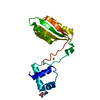
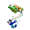
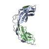
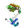
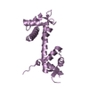
ANALYTICAL SIZE EXCLUSION CHROMATOGRPAHY SUPPORTS THE ASSIGNMENT OF A MONOMER AS A SIGNIFICANT OLIGOMERIZATION STATE IN SOLUTION. HOWEVER, CRYSTAL PACKING ANALYSIS SUGGESTS THAT THE PROTEIN HAS ASSOCIATED INTO TRIMERS THAT ARE PREDICTED TO BE STABLE.

-
Components

#1: Protein Tyrosine-protein kinase Lck / Leukocyte C-terminal Src kinase / LSK / Lymphocyte cell-specific protein-tyrosine kinase / Protein ...Leukocyte C-terminal Src kinase / LSK / Lymphocyte cell-specific protein-tyrosine kinase / Protein YT16 / Proto-oncogene Lck / T cell-specific protein-tyrosine kinase / p56-LCK


Mass: 19586.670 Da / Num. of mol.: 1 / Fragment: UNP residues 53-226
Source method: isolated from a genetically manipulated source
Source: (gene. exp.) Homo sapiens (human) / Gene: BC013200, LCK / Plasmid: SpeedET / Production host: Escherichia Coli (E. coli) / Strain (production host): HK100
References: UniProt: P06239, non-specific protein-tyrosine kinase
#2: Chemical ChemComp-SO4 / SULFATE ION


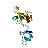
Mass: 96.063 Da / Num. of mol.: 1 / Source method: obtained synthetically / Formula: SO4
#3: Chemical ChemComp-GOL / GLYCEROL / GLYCERIN / PROPANE-1,2,3-TRIOL


Mass: 92.094 Da / Num. of mol.: 2 / Source method: obtained synthetically / Formula: C3H8O3
#4: Water ChemComp-HOH / water


Mass: 18.015 Da / Num. of mol.: 43 / Source method: isolated from a natural source / Formula: H2O
Sequence detailsTHIS CONSTRUCT WAS EXPRESSED WITH A PURIFICATION TAG MGSDKIHHHHHHENLYFQG. THE TAG WAS REMOVED WITH ...THIS CONSTRUCT WAS EXPRESSED WITH A PURIFICATION TAG MGSDKIHHHHHHENLYFQG. THE TAG WAS REMOVED WITH TEV PROTEASE LEAVING ONLY A GLYCINE (0) FOLLOWED BY RESIDUES 53-226 OF THE TARGET SEQUENCE.

-
Experimental details

-
Experiment

ExperimentMethod: X-RAY DIFFRACTION / Number of used crystals: 1

-
Sample preparation

CrystalDensity Matthews: 2.81 Å3/Da / Density % sol: 56.2 %
Crystal growTemperature: 277 K / Method: vapor diffusion, sitting drop / pH: 7.5
Details: 1.5M lithium sulfate, 0.1M HEPES pH 7.5, NANODROP, VAPOR DIFFUSION, SITTING DROP, temperature 277K

-
Data collection

DiffractionMean temperature: 100 K
Diffraction sourceSource: SYNCHROTRON / Site: ALS / Beamline: 8.2.2 / Wavelength: 0.9795
DetectorType: ADSC QUANTUM 315 / Detector: CCD / Date: Oct 16, 2011 / Details: KOHZU: Double Crystal Si(111)
RadiationMonochromator: Double Crystal Si(111) / Protocol: SINGLE WAVELENGTH / Monochromatic (M) / Laue (L): M / Scattering type: x-ray
Radiation wavelengthWavelength: 0.9795 Å / Relative weight: 1
ReflectionResolution: 2.36→29.834 Å / Num. all: 9434 / Num. obs: 9434 / % possible obs: 99.9 % / Redundancy: 4.9 % / Rsym value: 0.129 / Net I/σ(I): 9.3
Reflection shell

Diffraction-ID: 1

Resolution (Å)Redundancy (%)Rmerge(I) obsMean I/σ(I) obsNum. measured allNum. unique allRsym value% possible all
2.36-2.424.80.8650.932546740.865100
2.42-2.494.80.812132226680.81299.5
2.49-2.564.90.5921.231676430.59299.9
2.56-2.644.90.4811.631486360.48199.8
2.64-2.7350.4651.630356120.465100
2.73-2.8250.3422.229305820.342100
2.82-2.9350.332.429365860.33100
2.93-3.0550.2333.326775390.233100
3.05-3.184.90.174.426835430.17100
3.18-3.344.90.122624875070.122100
3.34-3.524.90.1166.424094960.116100
3.52-3.734.80.0897.822064620.089100
3.73-3.994.80.0739.520784370.073100
3.99-4.314.70.06110.618974030.061100
4.31-4.724.30.05612.616583840.056100
4.72-5.284.70.05212.616353490.052100
5.28-6.094.90.0621114963030.062100
6.09-7.464.90.0621113012680.062100
7.46-10.554.70.02923.510212150.029100
10.55-29.8344.10.02423.95271270.02495.5

-
Phasing

PhasingMethod: molecular replacement

-
Processing

Software
NameVersionClassificationNB
MolProbity3beta29model building
PDB_EXTRACT3.1data extraction
PHASER2.3.0phasing
SCALA3.3.20data scaling
PHENIX1.7.2refinement
MOSFLMdata reduction
RefinementMethod to determine structure: MOLECULAR REPLACEMENT / Resolution: 2.36→29.834 Å / Occupancy max: 1 / Occupancy min: 0.47 / SU ML: 0.61 / σ(F): 1.35 / Phase error: 26.18 / Stereochemistry target values: ML
Details: 1. ATOM RECORD CONTAINS SUM OF TLS AND RESIDUAL B FACTORS. 2. ANISOU RECORD CONTAINS SUM OF TLS AND RESIDUAL U FACTORS. 3. WATERS WERE EXCLUDED FROM AUTOMATIC TLS ASSIGNMENT. 4. SULFATE (SO4) ...Details: 1. ATOM RECORD CONTAINS SUM OF TLS AND RESIDUAL B FACTORS. 2. ANISOU RECORD CONTAINS SUM OF TLS AND RESIDUAL U FACTORS. 3. WATERS WERE EXCLUDED FROM AUTOMATIC TLS ASSIGNMENT. 4. SULFATE (SO4) FROM THE CRYSTALLIZATION AND GLYCEROL (GOL) USED AS A CRYOPROTECTANT WERE MODELED INTO THE STRUCTURE.
RfactorNum. reflection% reflection
Rfree0.2513 450 4.78 %
Rwork0.1911 --
obs0.194 9408 99.84 %
Solvent computationShrinkage radii: 0.9 Å / VDW probe radii: 1.11 Å / Solvent model: FLAT BULK SOLVENT MODEL / Bsol: 36.227 Å2 / ksol: 0.376 e/Å3
Displacement parametersBiso max: 130.39 Å2 / Biso mean: 47.5713 Å2 / Biso min: 8.07 Å2
Baniso -1Baniso -2Baniso -3
1-6.0335 Å2-0 Å20 Å2
2--6.0335 Å20 Å2
3----12.067 Å2
Refinement stepCycle: LAST / Resolution: 2.36→29.834 Å
ProteinNucleic acidLigandSolventTotal
Num. atoms1297 0 17 43 1357
Refine LS restraints
Refine-IDTypeDev idealNumber
X-RAY DIFFRACTIONf_bond_d0.0081349
X-RAY DIFFRACTIONf_angle_d1.1761826
X-RAY DIFFRACTIONf_chiral_restr0.071187
X-RAY DIFFRACTIONf_plane_restr0.005243
X-RAY DIFFRACTIONf_dihedral_angle_d14.718490
LS refinement shell

Refine-ID: X-RAY DIFFRACTION / Total num. of bins used: 3 / % reflection obs: 100 %

Resolution (Å)Rfactor RfreeNum. reflection RfreeRfactor RworkNum. reflection RworkNum. reflection all
2.3601-2.70140.33341500.260829153065
2.7014-3.40270.28641560.207729503106
3.4027-29.83660.20731440.163130933237
Refinement TLS params.

Method: refined / Refine-ID: X-RAY DIFFRACTION

IDL112)L122)L132)L222)L232)L332)S11 (Å °)S12 (Å °)S13 (Å °)S21 (Å °)S22 (Å °)S23 (Å °)S31 (Å °)S32 (Å °)S33 (Å °)T112)T122)T132)T222)T232)T332)Origin x (Å)Origin y (Å)Origin z (Å)
12.47570.1412-0.64642.88420.60913.1537-0.0799-0.52990.8110.72850.0334-0.3454-0.93080.7727-0.08350.6104-0.1171-0.12290.3416-0.06430.360859.1958-7.190611.3458
20.7298-0.1586-0.14361.9025-0.57954.1716-0.01240.0875-0.0908-0.27530.0775-0.07010.4703-0.083-0.06040.1743-0.0196-0.00050.1018-0.01190.109829.9311-18.995521.1831
34.881-0.8357-0.53914.74560.06084.22630.0544-0.01010.26450.0477-0.05560.0422-0.1716-0.1612-0.00110.0939-0.0043-0.01510.0776-0.05330.077129.4964-7.029822.9802
Refinement TLS group
IDRefine-IDRefine TLS-IDSelection detailsAuth asym-IDAuth seq-ID
1X-RAY DIFFRACTION1chain 'A' and (resseq 64:118)A0
2X-RAY DIFFRACTION2chain 'A' and (resseq 119:183)A0
3X-RAY DIFFRACTION3chain 'A' and (resseq 184:226)A0

+
About Yorodumi

-
News

-
Feb 9, 2022. New format data for meta-information of EMDB entries

New format data for meta-information of EMDB entries

  • Version 3 of the EMDB header file is now the official format.
  • The previous official version 1.9 will be removed from the archive.

Related info.:EMDB header

External links:wwPDB to switch to version 3 of the EMDB data model

-
Aug 12, 2020. Covid-19 info

Covid-19 info

URL: https://pdbjlvh1.pdbj.org/emnavi/covid19.php

New page: Covid-19 featured information page in EM Navigator.

Related info.:Covid-19 info / Mar 5, 2020. Novel coronavirus structure data

+
Mar 5, 2020. Novel coronavirus structure data

Novel coronavirus structure data

Related info.:Yorodumi Speices / Aug 12, 2020. Covid-19 info

External links:COVID-19 featured content - PDBj / Molecule of the Month (242):Coronavirus Proteases

+
Jan 31, 2019. EMDB accession codes are about to change! (news from PDBe EMDB page)

EMDB accession codes are about to change! (news from PDBe EMDB page)

  • The allocation of 4 digits for EMDB accession codes will soon come to an end. Whilst these codes will remain in use, new EMDB accession codes will include an additional digit and will expand incrementally as the available range of codes is exhausted. The current 4-digit format prefixed with “EMD-” (i.e. EMD-XXXX) will advance to a 5-digit format (i.e. EMD-XXXXX), and so on. It is currently estimated that the 4-digit codes will be depleted around Spring 2019, at which point the 5-digit format will come into force.
  • The EM Navigator/Yorodumi systems omit the EMD- prefix.

Related info.:Q: What is EMD? / ID/Accession-code notation in Yorodumi/EM Navigator

External links:EMDB Accession Codes are Changing Soon! / Contact to PDBj

+
Jul 12, 2017. Major update of PDB

Major update of PDB

  • wwPDB released updated PDB data conforming to the new PDBx/mmCIF dictionary.
  • This is a major update changing the version number from 4 to 5, and with Remediation, in which all the entries are updated.
  • In this update, many items about electron microscopy experimental information are reorganized (e.g. em_software).
  • Now, EM Navigator and Yorodumi are based on the updated data.

External links:wwPDB Remediation / Enriched Model Files Conforming to OneDep Data Standards Now Available in the PDB FTP Archive

-
Yorodumi

Thousand views of thousand structures

  • Yorodumi is a browser for structure data from EMDB, PDB, SASBDB, etc.
  • This page is also the successor to EM Navigator detail page, and also detail information page/front-end page for Omokage search.
  • The word "yorodu" (or yorozu) is an old Japanese word meaning "ten thousand". "mi" (miru) is to see.

Related info.:EMDB / PDB / SASBDB / Comparison of 3 databanks / Yorodumi Search / Aug 31, 2016. New EM Navigator & Yorodumi / Yorodumi Papers / Jmol/JSmol / Function and homology information / Changes in new EM Navigator and Yorodumi

Read more