 Movie
Movie Controller

[English] 日本語
 Yorodumi
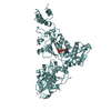
- PDB-5myb: Homodimerization of Tie2 Fibronectin-like domains 2 and 3 in spac... -
+ Open data

- Basic information
| Entry | Database: PDB / ID: 5myb | |||||||||
|---|---|---|---|---|---|---|---|---|---|---|
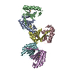
| Title | Homodimerization of Tie2 Fibronectin-like domains 2 and 3 in space group P21 | |||||||||
 Components Components | Angiopoietin-1 receptor | |||||||||
 Keywords Keywords | SIGNALING PROTEIN / receptor / fibronectin-like domains / dimerization / homotypic interactions | |||||||||
| Function / homology |  Function and homology information Function and homology informationTie signaling pathway / glomerulus vasculature development / transmembrane receptor protein kinase activity / regulation of endothelial cell apoptotic process / regulation of establishment or maintenance of cell polarity / heart trabecula formation / definitive hemopoiesis / regulation of vascular permeability / sprouting angiogenesis / endothelial cell proliferation ...Tie signaling pathway / glomerulus vasculature development / transmembrane receptor protein kinase activity / regulation of endothelial cell apoptotic process / regulation of establishment or maintenance of cell polarity / heart trabecula formation / definitive hemopoiesis / regulation of vascular permeability / sprouting angiogenesis / endothelial cell proliferation / positive regulation of Rho protein signal transduction / microvillus / positive regulation of intracellular signal transduction / positive regulation of focal adhesion assembly / negative regulation of endothelial cell apoptotic process / positive regulation of Rac protein signal transduction / Tie2 Signaling / positive regulation of endothelial cell proliferation / transmembrane receptor protein tyrosine kinase activity / substrate adhesion-dependent cell spreading / positive regulation of endothelial cell migration / negative regulation of angiogenesis / cell surface receptor protein tyrosine kinase signaling pathway / basal plasma membrane / cellular response to mechanical stimulus / receptor protein-tyrosine kinase / negative regulation of inflammatory response / positive regulation of angiogenesis / cell-cell junction / cell-cell signaling / heart development / RAF/MAP kinase cascade / signaling receptor activity / angiogenesis / basolateral plasma membrane / protein kinase activity / positive regulation of ERK1 and ERK2 cascade / positive regulation of MAPK cascade / cell surface receptor signaling pathway / positive regulation of phosphatidylinositol 3-kinase/protein kinase B signal transduction / signaling receptor complex / apical plasma membrane / ciliary basal body / membrane raft / focal adhesion / centrosome / negative regulation of apoptotic process / cell surface / extracellular region / ATP binding / identical protein binding / plasma membrane Similarity search - Function | |||||||||
| Biological species |  Homo sapiens (human) Homo sapiens (human) | |||||||||
| Method |  X-RAY DIFFRACTION / SYNCHROTRON / MOLECULAR REPLACEMENT / Resolution: 2.6 Å X-RAY DIFFRACTION / SYNCHROTRON / MOLECULAR REPLACEMENT / Resolution: 2.6 Å | |||||||||
 Authors Authors | Leppanen, V.-M. / Saharinen, P. / Alitalo, K. | |||||||||
 Citation Citation | Journal: Proc. Natl. Acad. Sci. U.S.A. / Year: 2017 Title: Structural basis of Tie2 activation and Tie2/Tie1 heterodimerization. Authors: Leppanen, V.M. / Saharinen, P. / Alitalo, K. | |||||||||
| History |
|
- Structure visualization
| Structure viewer | Molecule: MolmilJmol/JSmol |
|---|
- Downloads & links
-Download
| PDBx/mmCIF format | 5myb.cif.gz | 175.6 KB | Display | PDBx/mmCIF format |
|---|---|---|---|---|
| PDB format | pdb5myb.ent.gz | 137.6 KB | Display | PDB format |
| PDBx/mmJSON format | 5myb.json.gz | Tree view | PDBx/mmJSON format | |
| Others |  Other downloads Other downloads |
-Validation report
| Arichive directory | https://data.pdbj.org/pub/pdb/validation_reports/my/5mybftp://data.pdbj.org/pub/pdb/validation_reports/my/5myb | HTTPS FTP |
|---|
-Related structure data


-Links
 PDBj
PDBj


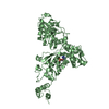
- Assembly
| Deposited unit | 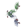
| ||||||||
|---|---|---|---|---|---|---|---|---|---|
| 1 |
| ||||||||
| Unit cell |
|
-Components
| #1: Protein | Mass: 37583.312 Da / Num. of mol.: 2 Source method: isolated from a genetically manipulated source Details: In addition to the Tie2 Fn-like domains, the entity includes mellitin signal peptide, a N-terminal cloning artefact (ADP), C-terminal Factor Xa cleavage site (IEGR) and His-tag. The first Fn- ...Details: In addition to the Tie2 Fn-like domains, the entity includes mellitin signal peptide, a N-terminal cloning artefact (ADP), C-terminal Factor Xa cleavage site (IEGR) and His-tag. The first Fn-like domain was not in the crystals apparently due to proteolytic removal prior crystallization. Source: (gene. exp.) Homo sapiens (human) / Gene: TEK, TIE2, VMCM, VMCM1Production host:  Spodoptera aff. frugiperda 2 RZ-2014 (butterflies/moths) Spodoptera aff. frugiperda 2 RZ-2014 (butterflies/moths)References: UniProt: Q02763, receptor protein-tyrosine kinase #2: Polysaccharide | beta-D-mannopyranose-(1-3)-2-acetamido-2-deoxy-beta-D-glucopyranose-(1-4)-2-acetamido-2-deoxy-beta- ...beta-D-mannopyranose-(1-3)-2-acetamido-2-deoxy-beta-D-glucopyranose-(1-4)-2-acetamido-2-deoxy-beta-D-glucopyranose | Source method: isolated from a genetically manipulated source #3: Polysaccharide | 2-acetamido-2-deoxy-beta-D-glucopyranose-(1-4)-2-acetamido-2-deoxy-beta-D-glucopyranose | Source method: isolated from a genetically manipulated source #4: Sugar | ChemComp-NAG /   Type: D-saccharide, beta linking / Mass: 221.208 Da / Num. of mol.: 4 Type: D-saccharide, beta linking / Mass: 221.208 Da / Num. of mol.: 4Source method: isolated from a genetically manipulated source Formula: C8H15NO6 #5: Water | ChemComp-HOH / |  Mass: 18.015 Da / Num. of mol.: 62 / Source method: isolated from a natural source / Formula: H2O Mass: 18.015 Da / Num. of mol.: 62 / Source method: isolated from a natural source / Formula: H2OHas protein modification | Y | |
|---|
-Experimental details
-Experiment
| Experiment | Method: X-RAY DIFFRACTION / Number of used crystals: 1 |
|---|
- Sample preparation
| Crystal | Density Matthews: 2.05 Å3/Da / Density % sol: 39.9 % / Description: rod-like |
|---|---|
| Crystal grow | Temperature: 294 K / Method: vapor diffusion, sitting drop / pH: 8 Details: 0.1 M Tris buffer at pH 7.0 - 8.5 and 14-20% PEG 3350 (w/v) Temp details: Room temperature |
-Data collection
| Diffraction | Mean temperature: 100 K / Ambient temp details: Liquid nitrogen cooling system |
|---|---|
| Diffraction source | Source: SYNCHROTRON / Site: ESRF  / Beamline: ID23-1 / Wavelength: 0.9762 Å / Beamline: ID23-1 / Wavelength: 0.9762 Å |
| Detector | Type: DECTRIS PILATUS 6M / Detector: PIXEL / Date: Feb 9, 2014 |
| Radiation | Monochromator: horizontally side diffracting Silicon 111 crystal Protocol: SINGLE WAVELENGTH / Monochromatic (M) / Laue (L): M / Scattering type: x-ray |
| Radiation wavelength | Wavelength: 0.9762 Å / Relative weight: 1 |
| Reflection | Biso Wilson estimate: 104.1 Å2 |
| Reflection shell | Resolution: 2.6→2.76 Å / Redundancy: 5.5 % / Mean I/σ(I) obs: 1.8 / Rsym value: 0.873 / % possible all: 93.6 |
- Processing
| Software |
| |||||||||||||||||||||||||||||||||||||||||||||||||||||||||||||||||||||||||||||||||||||||||||||||||||||||||||||||||||||||||||||||||||||||||||||||||||||||||||||||||||||||||||||||||||||||||||||||||||||||||||||||||||||||||||||||||
|---|---|---|---|---|---|---|---|---|---|---|---|---|---|---|---|---|---|---|---|---|---|---|---|---|---|---|---|---|---|---|---|---|---|---|---|---|---|---|---|---|---|---|---|---|---|---|---|---|---|---|---|---|---|---|---|---|---|---|---|---|---|---|---|---|---|---|---|---|---|---|---|---|---|---|---|---|---|---|---|---|---|---|---|---|---|---|---|---|---|---|---|---|---|---|---|---|---|---|---|---|---|---|---|---|---|---|---|---|---|---|---|---|---|---|---|---|---|---|---|---|---|---|---|---|---|---|---|---|---|---|---|---|---|---|---|---|---|---|---|---|---|---|---|---|---|---|---|---|---|---|---|---|---|---|---|---|---|---|---|---|---|---|---|---|---|---|---|---|---|---|---|---|---|---|---|---|---|---|---|---|---|---|---|---|---|---|---|---|---|---|---|---|---|---|---|---|---|---|---|---|---|---|---|---|---|---|---|---|---|---|---|---|---|---|---|---|---|---|---|---|---|---|---|---|---|---|
| Refinement | Method to determine structure: MOLECULAR REPLACEMENT Starting model: CRYSTAL STRUCTURE OF TIE2 FN-LIKE DOMAINS IN SPACE GROUP C2 Resolution: 2.6→39.8 Å / SU ML: 0.4 / Cross valid method: FREE R-VALUE / σ(F): 1.36 / Phase error: 33.66
| |||||||||||||||||||||||||||||||||||||||||||||||||||||||||||||||||||||||||||||||||||||||||||||||||||||||||||||||||||||||||||||||||||||||||||||||||||||||||||||||||||||||||||||||||||||||||||||||||||||||||||||||||||||||||||||||||
| Solvent computation | Shrinkage radii: 0.9 Å / VDW probe radii: 1.11 Å | |||||||||||||||||||||||||||||||||||||||||||||||||||||||||||||||||||||||||||||||||||||||||||||||||||||||||||||||||||||||||||||||||||||||||||||||||||||||||||||||||||||||||||||||||||||||||||||||||||||||||||||||||||||||||||||||||
| Refinement step | Cycle: LAST / Resolution: 2.6→39.8 Å
| |||||||||||||||||||||||||||||||||||||||||||||||||||||||||||||||||||||||||||||||||||||||||||||||||||||||||||||||||||||||||||||||||||||||||||||||||||||||||||||||||||||||||||||||||||||||||||||||||||||||||||||||||||||||||||||||||
| Refine LS restraints |
| |||||||||||||||||||||||||||||||||||||||||||||||||||||||||||||||||||||||||||||||||||||||||||||||||||||||||||||||||||||||||||||||||||||||||||||||||||||||||||||||||||||||||||||||||||||||||||||||||||||||||||||||||||||||||||||||||
| LS refinement shell |
| |||||||||||||||||||||||||||||||||||||||||||||||||||||||||||||||||||||||||||||||||||||||||||||||||||||||||||||||||||||||||||||||||||||||||||||||||||||||||||||||||||||||||||||||||||||||||||||||||||||||||||||||||||||||||||||||||
| Refinement TLS params. | Method: refined / Refine-ID: X-RAY DIFFRACTION
| |||||||||||||||||||||||||||||||||||||||||||||||||||||||||||||||||||||||||||||||||||||||||||||||||||||||||||||||||||||||||||||||||||||||||||||||||||||||||||||||||||||||||||||||||||||||||||||||||||||||||||||||||||||||||||||||||
| Refinement TLS group |
|
