 Movie
Movie Controller

[English] 日本語
 Yorodumi
- PDB-5mx8: Crystal structure of H. pylori purine nucleoside phosphorylase fr... -
+ Open data

- Basic information
| Entry | Database: PDB / ID: 5mx8 | ||||||
|---|---|---|---|---|---|---|---|
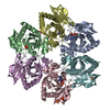
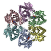
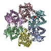
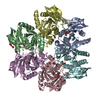
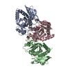
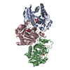
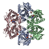
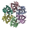
| Title | Crystal structure of H. pylori purine nucleoside phosphorylase from clinical isolate HpPNP-3 | ||||||
 Components Components | Purine nucleoside phosphorylase DeoD-type | ||||||
 Keywords Keywords | TRANSFERASE / purine nucleoside phosphorylase / clinical isolate / Helicobacter pylori / dead-end-complex | ||||||
| Function / homology |  Function and homology information Function and homology informationuridine phosphorylase / uridine phosphorylase activity / purine-nucleoside phosphorylase activity / purine-nucleoside phosphorylase / purine nucleoside catabolic process / cytosol Similarity search - Function | ||||||
| Biological species |   Helicobacter pylori (bacteria) Helicobacter pylori (bacteria) | ||||||
| Method |  X-RAY DIFFRACTION / MOLECULAR REPLACEMENT / Resolution: 2.4 Å X-RAY DIFFRACTION / MOLECULAR REPLACEMENT / Resolution: 2.4 Å | ||||||
 Authors Authors | Stefanic, Z. | ||||||
| Funding support |  Croatia, 1items Croatia, 1items
| ||||||
 Citation Citation | Journal: Int. J. Biol. Macromol. / Year: 2017 Title: Structural characterization of purine nucleoside phosphorylase from human pathogen Helicobacter pylori. Authors: Stefanic, Z. / Mikleusevic, G. / Luic, M. / Bzowska, A. / Lescic Asler, I. | ||||||
| History |
|
- Structure visualization
| Structure viewer | Molecule: MolmilJmol/JSmol |
|---|
- Downloads & links
-Download
| PDBx/mmCIF format | 5mx8.cif.gz | 64.3 KB | Display | PDBx/mmCIF format |
|---|---|---|---|---|
| PDB format | pdb5mx8.ent.gz | 47 KB | Display | PDB format |
| PDBx/mmJSON format | 5mx8.json.gz | Tree view | PDBx/mmJSON format | |
| Others |  Other downloads Other downloads |
-Validation report
| Arichive directory | https://data.pdbj.org/pub/pdb/validation_reports/mx/5mx8ftp://data.pdbj.org/pub/pdb/validation_reports/mx/5mx8 | HTTPS FTP |
|---|
-Related structure data
| Related structure data |  5mx4SC  5mx6C S: Starting model for refinement C: citing same article ( |
|---|---|
| Similar structure data |
-Links
 PDBj
PDBj
- Assembly
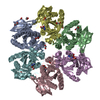
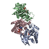
| Deposited unit | 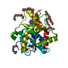
| ||||||||
|---|---|---|---|---|---|---|---|---|---|
| 1 | x 6
| ||||||||
| Unit cell |
|
-Components
| #1: Protein | Mass: 25817.096 Da / Num. of mol.: 1 / Source method: isolated from a natural source / Source: (natural) Helicobacter pylori (bacteria)References: UniProt: K2JXG0, UniProt: P56463*PLUS, purine-nucleoside phosphorylase | ||
|---|---|---|---|
| #2: Chemical | ChemComp-HPA /   Mass: 136.111 Da / Num. of mol.: 1 / Source method: obtained synthetically / Formula: C5H4N4O Mass: 136.111 Da / Num. of mol.: 1 / Source method: obtained synthetically / Formula: C5H4N4O | ||
| #3: Chemical | ChemComp-PO4 /   Mass: 94.971 Da / Num. of mol.: 1 / Source method: obtained synthetically / Formula: PO4 Mass: 94.971 Da / Num. of mol.: 1 / Source method: obtained synthetically / Formula: PO4 | ||
| #4: Chemical | ChemComp-PG4 /   Mass: 194.226 Da / Num. of mol.: 8 / Source method: obtained synthetically / Formula: C8H18O5 / Comment: precipitant*YM Mass: 194.226 Da / Num. of mol.: 8 / Source method: obtained synthetically / Formula: C8H18O5 / Comment: precipitant*YM#5: Water | ChemComp-HOH / |  Mass: 18.015 Da / Num. of mol.: 104 / Source method: isolated from a natural source / Formula: H2O Mass: 18.015 Da / Num. of mol.: 104 / Source method: isolated from a natural source / Formula: H2O |
-Experimental details
-Experiment
| Experiment | Method: X-RAY DIFFRACTION / Number of used crystals: 1 |
|---|
- Sample preparation
| Crystal | Density Matthews: 4.78 Å3/Da / Density % sol: 74.26 % |
|---|---|
| Crystal grow | Temperature: 291 K / Method: vapor diffusion, sitting drop / pH: 6.2 / Details: 0.2M NaCl, 0.1M Na/K phosphate pH 6.2, 50% PEG 200 |
-Data collection
| Diffraction | Mean temperature: 100 K |
|---|---|
| Diffraction source | Source: SEALED TUBE / Type: OXFORD DIFFRACTION ENHANCE ULTRA / Wavelength: 1.541 Å |
| Detector | Type: OXFORD RUBY CCD / Detector: CCD / Date: May 6, 2015 |
| Radiation | Protocol: SINGLE WAVELENGTH / Monochromatic (M) / Laue (L): M / Scattering type: x-ray |
| Radiation wavelength | Wavelength: 1.541 Å / Relative weight: 1 |
| Reflection | Resolution: 2.4→29.311 Å / Num. obs: 20243 / % possible obs: 99.4 % / Redundancy: 14 % / Net I/σ(I): 15.2 |
- Processing
| Software |
| ||||||||||||||||||||||||||||||||||||||||||||||||||||||||
|---|---|---|---|---|---|---|---|---|---|---|---|---|---|---|---|---|---|---|---|---|---|---|---|---|---|---|---|---|---|---|---|---|---|---|---|---|---|---|---|---|---|---|---|---|---|---|---|---|---|---|---|---|---|---|---|---|---|
| Refinement | Method to determine structure: MOLECULAR REPLACEMENT Starting model: 5MX4 Resolution: 2.4→29.311 Å / SU ML: 0.3 / Cross valid method: FREE R-VALUE / σ(F): 1.33 / Phase error: 25.37
| ||||||||||||||||||||||||||||||||||||||||||||||||||||||||
| Solvent computation | Shrinkage radii: 0.9 Å / VDW probe radii: 1.11 Å | ||||||||||||||||||||||||||||||||||||||||||||||||||||||||
| Displacement parameters | Biso max: 87.85 Å2 / Biso mean: 37.169 Å2 / Biso min: 19.09 Å2 | ||||||||||||||||||||||||||||||||||||||||||||||||||||||||
| Refinement step | Cycle: final / Resolution: 2.4→29.311 Å
| ||||||||||||||||||||||||||||||||||||||||||||||||||||||||
| Refine LS restraints |
| ||||||||||||||||||||||||||||||||||||||||||||||||||||||||
| LS refinement shell | Refine-ID: X-RAY DIFFRACTION / Total num. of bins used: 7
|
