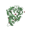
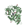
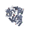
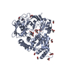
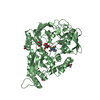
[English] 日本語
Yorodumi
- PDB-5mwu: Crystal structure of the periplasmic nickel-binding protein NikA ... -

+
Open data


ID or keywords:

Loading...

-
Basic information

Entry
Database: PDB / ID: 5mwu
TitleCrystal structure of the periplasmic nickel-binding protein NikA from Escherichia coli in complex with Ru(bpza)(CO)2Cl
ComponentsNickel-binding periplasmic protein
KeywordsMETAL BINDING PROTEIN / Artificial metalloenzymes / chlorine insertion / chlorhydrins / bio-inspired chemistry
Function / homology
Function and homology information


nickel cation import across plasma membrane / metal cluster binding / peptide transport / peptide transmembrane transporter activity / negative chemotaxis / nickel cation binding / transition metal ion binding / ATP-binding cassette (ABC) transporter complex, substrate-binding subunit-containing / outer membrane-bounded periplasmic space / periplasmic space ...nickel cation import across plasma membrane / metal cluster binding / peptide transport / peptide transmembrane transporter activity / negative chemotaxis / nickel cation binding / transition metal ion binding / ATP-binding cassette (ABC) transporter complex, substrate-binding subunit-containing / outer membrane-bounded periplasmic space / periplasmic space / heme binding / membrane
Similarity search - Function
Nickel ABC transporter, substrate-binding protein NikA / Solute-binding protein family 5, conserved site / Bacterial extracellular solute-binding proteins, family 5 signature. / Dipeptide-binding Protein; domain 3 / Dipeptide-binding Protein; Domain 3 / Peptide/nickel binding protein, MppA-type / Solute-binding protein family 5 domain / Solute-binding protein family 5 / Bacterial extracellular solute-binding proteins, family 5 Middle / Periplasmic binding protein-like II ...Nickel ABC transporter, substrate-binding protein NikA / Solute-binding protein family 5, conserved site / Bacterial extracellular solute-binding proteins, family 5 signature. / Dipeptide-binding Protein; domain 3 / Dipeptide-binding Protein; Domain 3 / Peptide/nickel binding protein, MppA-type / Solute-binding protein family 5 domain / Solute-binding protein family 5 / Bacterial extracellular solute-binding proteins, family 5 Middle / Periplasmic binding protein-like II / D-Maltodextrin-Binding Protein; domain 2 / Roll / 3-Layer(aba) Sandwich / Alpha Beta
Similarity search - Domain/homology
bis(pyrzol-1-yl)acetate scorpionate / ACETATE ION / CARBON MONOXIDE / Chem-EDT / : / RUTHENIUM ION / Nickel-binding periplasmic protein
Similarity search - Component
Biological speciesEscherichia coli K12 (bacteria)
MethodX-RAY DIFFRACTION / SYNCHROTRON / MOLECULAR REPLACEMENT / Resolution: 1.8 Å
AuthorsCavazza, C. / Lopez, S. / Rondot, L. / Iannello, M. / Boeri-Erba, E. / Burzlaff, N. / Strinitz, F. / Jorge-Robin, A. / Marchi-Delapierre, C. / Menage, S.
Funding support France, 1items
OrganizationGrant numberCountry
French National Research AgencyANR-14-CE06-0005-01 France
CitationJournal: To Be Published
Title: Efficient conversion of alkenes to chlorohydrins by a Ru-based artificial enzyme
Authors: Lopez, S. / Rondot, L. / Cavazza, C. / Iannello, M. / Boeri-Erba, E. / Burzlaff, N. / Strinitz, N. / Jorge-Robin, A. / Marchi-Delapierre, C. / Menage, S.
History
DepositionJan 20, 2017Deposition site: PDBE / Processing site: PDBE
Revision 1.0Feb 22, 2017Provider: repository / Type: Initial release
Revision 1.1Sep 6, 2017Group: Author supporting evidence / Category: pdbx_audit_support / Item: _pdbx_audit_support.funding_organization
Revision 1.2Nov 21, 2018Group: Data collection / Category: reflns / Item: _reflns.pdbx_Rsym_value
Revision 1.3Oct 16, 2019Group: Data collection / Category: reflns_shell
Revision 1.4Jan 17, 2024Group: Data collection / Database references / Refinement description
Category: chem_comp_atom / chem_comp_bond ...chem_comp_atom / chem_comp_bond / database_2 / pdbx_initial_refinement_model
Item: _database_2.pdbx_DOI / _database_2.pdbx_database_accession

-
Structure visualization

Structure viewerMolecule:
MolmilJmol/JSmol

Downloads & links

-
Assembly

Deposited unit
A: Nickel-binding periplasmic protein
B: Nickel-binding periplasmic protein
hetero molecules


Theoretical massNumber of molelcules
Total (without water)115,28133
Polymers112,7212
Non-polymers2,56031
Water11,998666
1
A: Nickel-binding periplasmic protein
hetero molecules


Theoretical massNumber of molelcules
Total (without water)57,86318
Polymers56,3611
Non-polymers1,50217
Water181
TypeNameSymmetry operationNumber
identity operation1_555x,y,z1
MethodPISA
2
B: Nickel-binding periplasmic protein
hetero molecules


Theoretical massNumber of molelcules
Total (without water)57,41915
Polymers56,3611
Non-polymers1,05814
Water181
TypeNameSymmetry operationNumber
identity operation1_555x,y,z1
MethodPISA
Unit cell
Length a, b, c (Å)86.303, 93.606, 124.211
Angle α, β, γ (deg.)90.00, 90.00, 90.00
Int Tables number19
Space group name H-MP212121

-
Components

-
Protein , 1 types, 2 molecules AB

#1: Protein Nickel-binding periplasmic protein


Mass: 56360.734 Da / Num. of mol.: 2
Source method: isolated from a genetically manipulated source
Source: (gene. exp.) Escherichia coli K12 (bacteria) / Gene: nikA, b3476, JW3441 / Production host: Escherichia coli BL21(DE3) (bacteria) / References: UniProt: P33590

-
Non-polymers , 10 types, 697 molecules

#2: Chemical
ChemComp-ACT / ACETATE ION / Acetate


Mass: 59.044 Da / Num. of mol.: 10 / Source method: obtained synthetically / Formula: C2H3O2
#3: Chemical ChemComp-SO4 / SULFATE ION / Sulfate


Mass: 96.063 Da / Num. of mol.: 1 / Source method: obtained synthetically / Formula: SO4
#4: Chemical
ChemComp-GOL / GLYCEROL / GLYCERIN / PROPANE-1,2,3-TRIOL / Glycerol


Mass: 92.094 Da / Num. of mol.: 12 / Source method: obtained synthetically / Formula: C3H8O3
#5: Chemical ChemComp-FE / FE (III) ION / Iron


Mass: 55.845 Da / Num. of mol.: 1 / Source method: obtained synthetically / Formula: Fe
#6: Chemical ChemComp-EDT / {[-(BIS-CARBOXYMETHYL-AMINO)-ETHYL]-CARBOXYMETHYL-AMINO}-ACETIC ACID / Ethylenediaminetetraacetic acid


Mass: 292.243 Da / Num. of mol.: 1 / Source method: obtained synthetically / Formula: C10H16N2O8
#7: Chemical ChemComp-RU / RUTHENIUM ION / Ruthenium


Mass: 101.070 Da / Num. of mol.: 1 / Source method: obtained synthetically / Formula: Ru
#8: Chemical ChemComp-CL / CHLORIDE ION / Chloride


Mass: 35.453 Da / Num. of mol.: 2 / Source method: obtained synthetically / Formula: Cl
#9: Chemical ChemComp-6RP / bis(pyrzol-1-yl)acetate scorpionate / BPZ


Mass: 192.175 Da / Num. of mol.: 1 / Source method: obtained synthetically / Formula: C8H8N4O2
#10: Chemical ChemComp-CMO / CARBON MONOXIDE / Carbon monoxide


Mass: 28.010 Da / Num. of mol.: 2 / Source method: obtained synthetically / Formula: CO
#11: Water ChemComp-HOH / water / Water


Mass: 18.015 Da / Num. of mol.: 666 / Source method: isolated from a natural source / Formula: H2O

-
Experimental details

-
Experiment

ExperimentMethod: X-RAY DIFFRACTION / Number of used crystals: 1

-
Sample preparation

CrystalDensity Matthews: 2.23 Å3/Da / Density % sol: 44.73 %
Crystal growTemperature: 293 K / Method: vapor diffusion, hanging drop / pH: 4.7
Details: 15 mg/ml protei solution mixed with 1.8 M ammonium sulfate, 100 mM sodium acetate pH 4.7

-
Data collection

DiffractionMean temperature: 93 K
Diffraction sourceSource: SYNCHROTRON / Site: ESRF / Beamline: BM30A / Wavelength: 0.979769 Å
DetectorType: ADSC QUANTUM 315r / Detector: CCD / Date: Feb 28, 2015
RadiationProtocol: SINGLE WAVELENGTH / Monochromatic (M) / Laue (L): M / Scattering type: x-ray
Radiation wavelengthWavelength: 0.979769 Å / Relative weight: 1
ReflectionResolution: 1.8→46.8 Å / Num. obs: 92563 / % possible obs: 98.8 % / Redundancy: 4.98 % / Biso Wilson estimate: 27.6 Å2 / Rsym value: 0.057 / Net I/σ(I): 21.24

-
Processing

Software
NameVersionClassification
REFMAC5.8.0049refinement
XDSdata reduction
XDSdata scaling
PHENIXphasing
RefinementMethod to determine structure: MOLECULAR REPLACEMENT
Starting model: 1ZLQ
Resolution: 1.8→46.8 Å / Cor.coef. Fo:Fc: 0.966 / Cor.coef. Fo:Fc free: 0.949 / SU B: 3.145 / SU ML: 0.093 / Cross valid method: THROUGHOUT / ESU R: 0.126 / ESU R Free: 0.121 / Details: HYDROGENS HAVE BEEN ADDED IN THE RIDING POSITIONS
RfactorNum. reflection% reflectionSelection details
Rfree0.20818 4629 5 %RANDOM
Rwork0.16769 ---
obs0.16972 87934 98.77 %-
Solvent computationIon probe radii: 0.8 Å / Shrinkage radii: 0.8 Å / VDW probe radii: 1.2 Å
Displacement parametersBiso mean: 27.073 Å2
Baniso -1Baniso -2Baniso -3
1-1.24 Å20 Å20 Å2
2---1.05 Å20 Å2
3----0.19 Å2
Refinement stepCycle: 1 / Resolution: 1.8→46.8 Å
ProteinNucleic acidLigandSolventTotal
Num. atoms7847 0 159 666 8672
Refine LS restraints
Refine-IDTypeDev idealDev ideal targetNumber
X-RAY DIFFRACTIONr_bond_refined_d0.020.0198369
X-RAY DIFFRACTIONr_bond_other_d0.0010.027874
X-RAY DIFFRACTIONr_angle_refined_deg1.8981.97211417
X-RAY DIFFRACTIONr_angle_other_deg2.347318166
X-RAY DIFFRACTIONr_dihedral_angle_1_deg6.60851052
X-RAY DIFFRACTIONr_dihedral_angle_2_deg38.32824.545385
X-RAY DIFFRACTIONr_dihedral_angle_3_deg12.213151347
X-RAY DIFFRACTIONr_dihedral_angle_4_deg16.4721545
X-RAY DIFFRACTIONr_chiral_restr0.1190.21257
X-RAY DIFFRACTIONr_gen_planes_refined0.0110.0219533
X-RAY DIFFRACTIONr_gen_planes_other0.0130.021896
X-RAY DIFFRACTIONr_nbd_refined
X-RAY DIFFRACTIONr_nbd_other
X-RAY DIFFRACTIONr_nbtor_refined
X-RAY DIFFRACTIONr_nbtor_other
X-RAY DIFFRACTIONr_xyhbond_nbd_refined
X-RAY DIFFRACTIONr_xyhbond_nbd_other
X-RAY DIFFRACTIONr_metal_ion_refined
X-RAY DIFFRACTIONr_metal_ion_other
X-RAY DIFFRACTIONr_symmetry_vdw_refined
X-RAY DIFFRACTIONr_symmetry_vdw_other
X-RAY DIFFRACTIONr_symmetry_hbond_refined
X-RAY DIFFRACTIONr_symmetry_hbond_other
X-RAY DIFFRACTIONr_symmetry_metal_ion_refined
X-RAY DIFFRACTIONr_symmetry_metal_ion_other
X-RAY DIFFRACTIONr_mcbond_it2.4342.4854059
X-RAY DIFFRACTIONr_mcbond_other2.4122.4814050
X-RAY DIFFRACTIONr_mcangle_it3.2243.7045069
X-RAY DIFFRACTIONr_mcangle_other3.2243.7045069
X-RAY DIFFRACTIONr_scbond_it3.3172.8184310
X-RAY DIFFRACTIONr_scbond_other3.3172.8184310
X-RAY DIFFRACTIONr_scangle_it
X-RAY DIFFRACTIONr_scangle_other4.8124.0956320
X-RAY DIFFRACTIONr_long_range_B_refined6.22821.16910098
X-RAY DIFFRACTIONr_long_range_B_other6.22921.16810098
X-RAY DIFFRACTIONr_rigid_bond_restr
X-RAY DIFFRACTIONr_sphericity_free
X-RAY DIFFRACTIONr_sphericity_bonded
LS refinement shellResolution: 1.8→1.847 Å / Total num. of bins used: 20
RfactorNum. reflection% reflection
Rfree0.328 340 -
Rwork0.274 6457 -
obs--99.68 %

+
About Yorodumi

-
News

-
Feb 9, 2022. New format data for meta-information of EMDB entries

New format data for meta-information of EMDB entries

  • Version 3 of the EMDB header file is now the official format.
  • The previous official version 1.9 will be removed from the archive.

Related info.:EMDB header

External links:wwPDB to switch to version 3 of the EMDB data model

-
Aug 12, 2020. Covid-19 info

Covid-19 info

URL: https://pdbj.org/emnavi/covid19.php

New page: Covid-19 featured information page in EM Navigator.

Related info.:Covid-19 info / Mar 5, 2020. Novel coronavirus structure data

+
Mar 5, 2020. Novel coronavirus structure data

Novel coronavirus structure data

Related info.:Yorodumi Speices / Aug 12, 2020. Covid-19 info

External links:COVID-19 featured content - PDBj / Molecule of the Month (242):Coronavirus Proteases

+
Jan 31, 2019. EMDB accession codes are about to change! (news from PDBe EMDB page)

EMDB accession codes are about to change! (news from PDBe EMDB page)

  • The allocation of 4 digits for EMDB accession codes will soon come to an end. Whilst these codes will remain in use, new EMDB accession codes will include an additional digit and will expand incrementally as the available range of codes is exhausted. The current 4-digit format prefixed with “EMD-” (i.e. EMD-XXXX) will advance to a 5-digit format (i.e. EMD-XXXXX), and so on. It is currently estimated that the 4-digit codes will be depleted around Spring 2019, at which point the 5-digit format will come into force.
  • The EM Navigator/Yorodumi systems omit the EMD- prefix.

Related info.:Q: What is EMD? / ID/Accession-code notation in Yorodumi/EM Navigator

External links:EMDB Accession Codes are Changing Soon! / Contact to PDBj

+
Jul 12, 2017. Major update of PDB

Major update of PDB

  • wwPDB released updated PDB data conforming to the new PDBx/mmCIF dictionary.
  • This is a major update changing the version number from 4 to 5, and with Remediation, in which all the entries are updated.
  • In this update, many items about electron microscopy experimental information are reorganized (e.g. em_software).
  • Now, EM Navigator and Yorodumi are based on the updated data.

External links:wwPDB Remediation / Enriched Model Files Conforming to OneDep Data Standards Now Available in the PDB FTP Archive

-
Yorodumi

Thousand views of thousand structures

  • Yorodumi is a browser for structure data from EMDB, PDB, SASBDB, etc.
  • This page is also the successor to EM Navigator detail page, and also detail information page/front-end page for Omokage search.
  • The word "yorodu" (or yorozu) is an old Japanese word meaning "ten thousand". "mi" (miru) is to see.

Related info.:EMDB / PDB / SASBDB / Comparison of 3 databanks / Yorodumi Search / Aug 31, 2016. New EM Navigator & Yorodumi / Yorodumi Papers / Jmol/JSmol / Function and homology information / Changes in new EM Navigator and Yorodumi

Read more