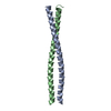
 Movie
Movie Controller


+ Open data

- Basic information
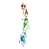
| Entry | Database: PDB / ID: 5mwb | |||||||||
|---|---|---|---|---|---|---|---|---|---|---|
| Title | Human Notch-2 EGF11-13 | |||||||||
 Components Components | Neurogenic locus notch homolog protein 2 | |||||||||
 Keywords Keywords | SIGNALING PROTEIN / EGF / Notch / receptor / signaling | |||||||||
| Function / homology |  Function and homology information Function and homology informationcholangiocyte proliferation / regulation of osteoclast development / intrahepatic bile duct development / glomerular capillary formation / ciliary body morphogenesis / podocyte development / proximal tubule development / Defective LFNG causes SCDO3 / marginal zone B cell differentiation / morphogenesis of an epithelial sheet ...cholangiocyte proliferation / regulation of osteoclast development / intrahepatic bile duct development / glomerular capillary formation / ciliary body morphogenesis / podocyte development / proximal tubule development / Defective LFNG causes SCDO3 / marginal zone B cell differentiation / morphogenesis of an epithelial sheet / Pre-NOTCH Processing in the Endoplasmic Reticulum / atrioventricular node development / cellular response to tumor cell / atrial septum morphogenesis / positive regulation of keratinocyte proliferation / hepatocyte proliferation / positive regulation of smooth muscle cell differentiation / left/right axis specification / NOTCH2 intracellular domain regulates transcription / Pre-NOTCH Processing in Golgi / pulmonary valve morphogenesis / placenta blood vessel development / myeloid dendritic cell differentiation / cell fate determination / bone remodeling / positive regulation of BMP signaling pathway / inflammatory response to antigenic stimulus / positive regulation of osteoclast differentiation / positive regulation of Ras protein signal transduction / NOTCH4 Intracellular Domain Regulates Transcription / embryonic limb morphogenesis / Notch-HLH transcription pathway / heart looping / humoral immune response / hemopoiesis / BMP signaling pathway / NF-kappaB binding / Notch signaling pathway / NOTCH2 Activation and Transmission of Signal to the Nucleus / axon guidance / animal organ morphogenesis / wound healing / positive regulation of miRNA transcription / Pre-NOTCH Transcription and Translation / multicellular organism growth / nervous system development / signaling receptor activity / in utero embryonic development / positive regulation of ERK1 and ERK2 cascade / transcription coactivator activity / signaling receptor complex / defense response to bacterium / intracellular signal transduction / cilium / positive regulation of apoptotic process / Golgi membrane / negative regulation of gene expression / apoptotic process / calcium ion binding / endoplasmic reticulum membrane / negative regulation of apoptotic process / enzyme binding / cell surface / negative regulation of transcription by RNA polymerase II / Golgi apparatus / positive regulation of transcription by RNA polymerase II / extracellular region / nucleoplasm / membrane / nucleus / plasma membrane Similarity search - Function | |||||||||
| Biological species |  Homo sapiens (human) Homo sapiens (human) | |||||||||
| Method |  X-RAY DIFFRACTION / SYNCHROTRON / MOLECULAR REPLACEMENT / Resolution: 1.86 Å X-RAY DIFFRACTION / SYNCHROTRON / MOLECULAR REPLACEMENT / Resolution: 1.86 Å | |||||||||
 Authors Authors | Suckling, R.J. / Handford, P.A. / Lea, S.M. | |||||||||
| Funding support |  United Kingdom, 1items United Kingdom, 1items
| |||||||||
 Citation Citation | Journal: EMBO J. / Year: 2017 Title: Structural and functional dissection of the interplay between lipid and Notch binding by human Notch ligands. Authors: Suckling, R.J. / Korona, B. / Whiteman, P. / Chillakuri, C. / Holt, L. / Handford, P.A. / Lea, S.M. | |||||||||
| History |
|
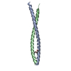
- Structure visualization
| Structure viewer | Molecule: MolmilJmol/JSmol |
|---|
- Downloads & links
-Download
| PDBx/mmCIF format | 5mwb.cif.gz | 45.7 KB | Display | PDBx/mmCIF format |
|---|---|---|---|---|
| PDB format | pdb5mwb.ent.gz | 28 KB | Display | PDB format |
| PDBx/mmJSON format | 5mwb.json.gz | Tree view | PDBx/mmJSON format | |
| Others |  Other downloads Other downloads |
-Validation report
| Arichive directory | https://data.pdbj.org/pub/pdb/validation_reports/mw/5mwbftp://data.pdbj.org/pub/pdb/validation_reports/mw/5mwb | HTTPS FTP |
|---|
-Related structure data
| Related structure data | 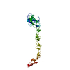 5mvxC 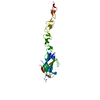 5mw5C 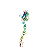 5mw7C  5mwfC  2vj3S S: Starting model for refinement C: citing same article ( |
|---|---|
| Similar structure data |

-Links
 PDBj
PDBj


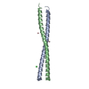
- Assembly
| Deposited unit | 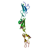
| ||||||||
|---|---|---|---|---|---|---|---|---|---|
| 1 |
| ||||||||
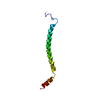
| Unit cell |
|
-Components
-Protein , 1 types, 1 molecules A
| #1: Protein | Mass: 17145.312 Da / Num. of mol.: 1 / Fragment: UNP residues 414-532 Source method: isolated from a genetically manipulated source Source: (gene. exp.) Homo sapiens (human) / Gene: NOTCH2 / Plasmid: pEXS2-2 / Cell (production host): Schneider 2 / Production host:  |
|---|
-Sugars , 3 types, 3 molecules 

| #2: Polysaccharide | beta-D-xylopyranose-(1-3)-beta-D-glucopyranose Source method: isolated from a genetically manipulated source |
|---|---|
| #3: Sugar | ChemComp-FUC /  Type: L-saccharide, alpha linking / Mass: 164.156 Da / Num. of mol.: 1 Type: L-saccharide, alpha linking / Mass: 164.156 Da / Num. of mol.: 1Source method: isolated from a genetically manipulated source Formula: C6H12O5 |
| #4: Sugar | ChemComp-BGC /  Type: D-saccharide, beta linking / Mass: 180.156 Da / Num. of mol.: 1 Type: D-saccharide, beta linking / Mass: 180.156 Da / Num. of mol.: 1Source method: isolated from a genetically manipulated source Formula: C6H12O6 |
-Non-polymers , 2 types, 124 molecules 

| #5: Chemical |  Mass: 40.078 Da / Num. of mol.: 3 / Source method: obtained synthetically / Formula: Ca Mass: 40.078 Da / Num. of mol.: 3 / Source method: obtained synthetically / Formula: Ca#6: Water | ChemComp-HOH / | Mass: 18.015 Da / Num. of mol.: 121 / Source method: isolated from a natural source / Formula: H2O |
|---|
-Details
| Has protein modification | Y |
|---|
-Experimental details
-Experiment
| Experiment | Method: X-RAY DIFFRACTION / Number of used crystals: 1 |
|---|
- Sample preparation
| Crystal | Density Matthews: 1.84 Å3/Da / Density % sol: 33.04 % |
|---|---|
| Crystal grow | Temperature: 293 K / Method: vapor diffusion / pH: 6.5 / Details: PEG 8000, Sodium acetate, Sodium cacodylate |
-Data collection
| Diffraction | Mean temperature: 100 K |
|---|---|
| Diffraction source | Source: SYNCHROTRON / Site: Diamond / Beamline: I04 / Wavelength: 0.98 Å |
| Detector | Type: DECTRIS PILATUS 6M-F / Detector: PIXEL / Date: Apr 19, 2015 |
| Radiation | Protocol: SINGLE WAVELENGTH / Monochromatic (M) / Laue (L): M / Scattering type: x-ray |
| Radiation wavelength | Wavelength: 0.98 Å / Relative weight: 1 |
| Reflection | Resolution: 1.86→41.8 Å / Num. obs: 11247 / % possible obs: 99.5 % / Redundancy: 5.2 % / CC1/2: 0.998 / Rmerge(I) obs: 0.077 / Net I/σ(I): 11.2 |
| Reflection shell | Resolution: 1.86→1.91 Å / Redundancy: 5.1 % / Rmerge(I) obs: 1.067 / Mean I/σ(I) obs: 1.3 / CC1/2: 0.524 / % possible all: 99.3 |
- Processing
| Software |
| |||||||||||||||||||||||||||||||||||
|---|---|---|---|---|---|---|---|---|---|---|---|---|---|---|---|---|---|---|---|---|---|---|---|---|---|---|---|---|---|---|---|---|---|---|---|---|
| Refinement | Method to determine structure: MOLECULAR REPLACEMENT Starting model: 2VJ3 Resolution: 1.86→39.003 Å / SU ML: 0.24 / Cross valid method: FREE R-VALUE / σ(F): 1.34 / Phase error: 33.94
| |||||||||||||||||||||||||||||||||||
| Solvent computation | Shrinkage radii: 0.9 Å / VDW probe radii: 1.11 Å | |||||||||||||||||||||||||||||||||||
| Refinement step | Cycle: LAST / Resolution: 1.86→39.003 Å
| |||||||||||||||||||||||||||||||||||
| Refine LS restraints |
| |||||||||||||||||||||||||||||||||||
| LS refinement shell |
|
