[English] 日本語
Yorodumi
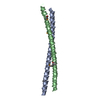
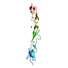
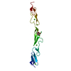
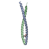
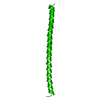
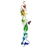
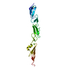
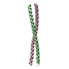
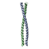
- PDB-2q6q: Crystal structure of Spc42p, a critical component of spindle pole... -

+
Open data


ID or keywords:

Loading...

-
Basic information

Entry
Database: PDB / ID: 2q6q
TitleCrystal structure of Spc42p, a critical component of spindle pole body in budding yeast
ComponentsSpindle pole body component SPC42
KeywordsCELL CYCLE / Spc42p / spindle pole body / budding yeast
Function / homology
Function and homology information


intermediate layer of spindle pole body / central plaque of spindle pole body / spindle pole body duplication / regulation of microtubule nucleation / structural constituent of cytoskeleton / nucleus / cytoplasm
Similarity search - Function
Spindle pole body component Spc42 / Spindle pole body component Spc42p / Geminin coiled-coil domain / Single alpha-helices involved in coiled-coils or other helix-helix interfaces / Up-down Bundle / Mainly Alpha
Similarity search - Domain/homology
: / Spindle pole body component SPC42
Similarity search - Component
Biological speciesSaccharomyces cerevisiae (brewer's yeast)
MethodX-RAY DIFFRACTION / SYNCHROTRON / MOLECULAR REPLACEMENT / Resolution: 1.97 Å
AuthorsMalashkevich, V.N. / Newman, J.R. / Kim, P.S.
CitationJournal: Biochemistry / Year: 2008
Title: Analysis of coiled-coil interactions between core proteins of the spindle pole body.
Authors: Zizlsperger, N. / Malashkevich, V.N. / Pillay, S. / Keating, A.E.
History
DepositionJun 5, 2007Deposition site: RCSB / Processing site: RCSB
Revision 1.0May 13, 2008Provider: repository / Type: Initial release
Revision 1.1Jul 13, 2011Group: Advisory / Version format compliance
Revision 1.2Feb 21, 2024Group: Data collection / Database references / Derived calculations
Category: chem_comp_atom / chem_comp_bond ...chem_comp_atom / chem_comp_bond / database_2 / struct_conn / struct_site
Item: _database_2.pdbx_DOI / _database_2.pdbx_database_accession ..._database_2.pdbx_DOI / _database_2.pdbx_database_accession / _struct_conn.pdbx_dist_value / _struct_conn.ptnr1_auth_asym_id / _struct_conn.ptnr1_auth_comp_id / _struct_conn.ptnr1_auth_seq_id / _struct_conn.ptnr1_label_asym_id / _struct_conn.ptnr1_label_atom_id / _struct_conn.ptnr1_label_comp_id / _struct_conn.ptnr1_label_seq_id / _struct_conn.ptnr2_auth_asym_id / _struct_conn.ptnr2_auth_comp_id / _struct_conn.ptnr2_auth_seq_id / _struct_conn.ptnr2_label_asym_id / _struct_conn.ptnr2_label_atom_id / _struct_conn.ptnr2_label_comp_id / _struct_conn.ptnr2_label_seq_id / _struct_site.pdbx_auth_asym_id / _struct_site.pdbx_auth_comp_id / _struct_site.pdbx_auth_seq_id
Revision 1.3Apr 3, 2024Group: Refinement description / Category: pdbx_initial_refinement_model

-
Structure visualization

Structure viewerMolecule:
MolmilJmol/JSmol

Downloads & links

-
Assembly

Deposited unit
A: Spindle pole body component SPC42
B: Spindle pole body component SPC42
hetero molecules


Theoretical massNumber of molelcules
Total (without water)17,9276
Polymers17,7142
Non-polymers2124
Water2,144119
1


  • Idetical with deposited unit
  • defined by author&software
TypeNameSymmetry operationNumber
identity operation1_555x,y,z1
Buried area3670 Å2
ΔGint-62.7 kcal/mol
Surface area9710 Å2
MethodPISA
Unit cell
Length a, b, c (Å)42.860, 57.790, 57.170
Angle α, β, γ (deg.)90.00, 90.00, 90.00
Int Tables number19
Space group name H-MP212121

-
Components

#1: Protein Spindle pole body component SPC42 /


Mass: 8857.232 Da / Num. of mol.: 2 / Fragment: residues 65-138
Source method: isolated from a genetically manipulated source
Source: (gene. exp.) Saccharomyces cerevisiae (brewer's yeast)
Gene: SPC42 / Plasmid: pAED4 / Production host: Escherichia coli (E. coli) / Strain (production host): BL21-pLysS / References: UniProt: P36094
#2: Chemical ChemComp-CO / COBALT (II) ION


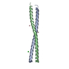
Mass: 58.933 Da / Num. of mol.: 3 / Source method: obtained synthetically / Formula: Co
#3: Chemical ChemComp-CL / CHLORIDE ION / Chloride


Mass: 35.453 Da / Num. of mol.: 1 / Source method: obtained synthetically / Formula: Cl
#4: Water ChemComp-HOH / water / Water


Mass: 18.015 Da / Num. of mol.: 119 / Source method: isolated from a natural source / Formula: H2O

-
Experimental details

-
Experiment

ExperimentMethod: X-RAY DIFFRACTION / Number of used crystals: 1

-
Sample preparation

CrystalDensity Matthews: 2 Å3/Da / Density % sol: 38.44 %
Crystal growTemperature: 294 K / Method: vapor diffusion, hanging drop / pH: 8.25
Details: 100 mM Tris, 200 mM cobalt chloride, 25% PEG 3350, pH 8.25, VAPOR DIFFUSION, HANGING DROP, temperature 294K

-
Data collection

DiffractionMean temperature: 100 K
Diffraction sourceSource: SYNCHROTRON / Site: NSLS / Beamline: X4A / Wavelength: 0.979 Å
DetectorType: ADSC QUANTUM 4 / Detector: CCD / Date: Mar 7, 2001 / Details: X4A
RadiationMonochromator: X4A / Protocol: SINGLE WAVELENGTH / Monochromatic (M) / Laue (L): M / Scattering type: x-ray
Radiation wavelengthWavelength: 0.979 Å / Relative weight: 1
ReflectionResolution: 1.97→35 Å / Num. all: 10537 / Num. obs: 9799 / % possible obs: 93.9 % / Redundancy: 4.5 % / Rmerge(I) obs: 0.075 / Rsym value: 0.075 / Net I/σ(I): 8.2
Reflection shellResolution: 1.97→2.02 Å / Redundancy: 3 % / Rmerge(I) obs: 0.37 / Mean I/σ(I) obs: 3.1 / Rsym value: 0.37 / % possible all: 47

-
Processing

Software
NameVersionClassification
REFMAC5.3.0034refinement
ADSCQuantumdata collection
DENZOdata reduction
SCALEPACKdata scaling
AMoREphasing
RefinementMethod to determine structure: MOLECULAR REPLACEMENT
Starting model: GCN4

Resolution: 1.97→18.36 Å / Cor.coef. Fo:Fc: 0.935 / Cor.coef. Fo:Fc free: 0.876 / SU B: 9.951 / SU ML: 0.146 / TLS residual ADP flag: LIKELY RESIDUAL / Cross valid method: THROUGHOUT / ESU R: 0.219 / ESU R Free: 0.216 / Stereochemistry target values: MAXIMUM LIKELIHOOD / Details: HYDROGENS HAVE BEEN ADDED IN THE RIDING POSITIONS
RfactorNum. reflection% reflectionSelection details
Rfree0.29655 475 4.8 %RANDOM
Rwork0.21163 ---
obs0.21565 9404 94.68 %-
all-10055 --
Solvent computationIon probe radii: 0.8 Å / Shrinkage radii: 0.8 Å / VDW probe radii: 1.2 Å / Solvent model: BABINET MODEL WITH MASK
Displacement parametersBiso mean: 36.772 Å2
Baniso -1Baniso -2Baniso -3
1-0.03 Å20 Å20 Å2
2--0 Å20 Å2
3----0.04 Å2
Refinement stepCycle: LAST / Resolution: 1.97→18.36 Å
ProteinNucleic acidLigandSolventTotal
Num. atoms1065 0 4 119 1188
Refine LS restraints
Refine-IDTypeDev idealDev ideal targetNumber
X-RAY DIFFRACTIONr_bond_refined_d0.0190.0221074
X-RAY DIFFRACTIONr_bond_other_d
X-RAY DIFFRACTIONr_angle_refined_deg1.9082.0041428
X-RAY DIFFRACTIONr_angle_other_deg
X-RAY DIFFRACTIONr_dihedral_angle_1_deg5.4845127
X-RAY DIFFRACTIONr_dihedral_angle_2_deg38.56227.45555
X-RAY DIFFRACTIONr_dihedral_angle_3_deg19.53215260
X-RAY DIFFRACTIONr_dihedral_angle_4_deg17.793154
X-RAY DIFFRACTIONr_chiral_restr0.1310.2166
X-RAY DIFFRACTIONr_gen_planes_refined0.0070.02748
X-RAY DIFFRACTIONr_gen_planes_other
X-RAY DIFFRACTIONr_nbd_refined0.2430.2503
X-RAY DIFFRACTIONr_nbd_other
X-RAY DIFFRACTIONr_nbtor_refined0.3030.2751
X-RAY DIFFRACTIONr_nbtor_other
X-RAY DIFFRACTIONr_xyhbond_nbd_refined0.2710.296
X-RAY DIFFRACTIONr_xyhbond_nbd_other
X-RAY DIFFRACTIONr_metal_ion_refined0.090.21
X-RAY DIFFRACTIONr_metal_ion_other
X-RAY DIFFRACTIONr_symmetry_vdw_refined0.2510.243
X-RAY DIFFRACTIONr_symmetry_vdw_other
X-RAY DIFFRACTIONr_symmetry_hbond_refined0.2660.218
X-RAY DIFFRACTIONr_symmetry_hbond_other
X-RAY DIFFRACTIONr_symmetry_metal_ion_refined
X-RAY DIFFRACTIONr_symmetry_metal_ion_other
X-RAY DIFFRACTIONr_mcbond_it1.8421.5636
X-RAY DIFFRACTIONr_mcbond_other
X-RAY DIFFRACTIONr_mcangle_it9.27501030
X-RAY DIFFRACTIONr_scbond_it20.77850438
X-RAY DIFFRACTIONr_scangle_it7.6074.5397
X-RAY DIFFRACTIONr_rigid_bond_restr
X-RAY DIFFRACTIONr_sphericity_free
X-RAY DIFFRACTIONr_sphericity_bonded
LS refinement shellResolution: 1.974→2.025 Å / Total num. of bins used: 20
RfactorNum. reflection% reflection
Rfree0.212 19 -
Rwork0.248 391 -
obs--55.18 %
Refinement TLS params.

Method: refined / Refine-ID: X-RAY DIFFRACTION

IDL112)L122)L132)L222)L232)L332)S11 (Å °)S12 (Å °)S13 (Å °)S21 (Å °)S22 (Å °)S23 (Å °)S31 (Å °)S32 (Å °)S33 (Å °)T112)T122)T132)T222)T232)T332)Origin x (Å)Origin y (Å)Origin z (Å)
16.4641.62333.6420.55961.07872.2291-0.0553-0.1149-0.0938-0.05770.0426-0.0204-0.042-0.03980.01270.012-0.026-0.0129-0.03320.0251-0.03434.52888.39589.9544
25.2387-0.05522.67090.08310.01581.46210.10070.0313-0.1053-0.0094-0.03110.02650.06480.0155-0.0696-0.0018-0.0057-0.0153-0.04920.0088-0.01495.46548.7185.1219
35.35590.1896.64090.05130.34188.4931-0.47210.28960.3483-0.06380.07510.0341-0.57270.38460.397-0.00650.00170.00420.0096-0.0013-0.00153.50289.764212.8767
Refinement TLS group
IDRefine-IDRefine TLS-IDAuth asym-IDLabel asym-IDAuth seq-IDLabel seq-ID
1X-RAY DIFFRACTION1AA67 - 1283 - 64
2X-RAY DIFFRACTION2BB67 - 1313 - 67
3X-RAY DIFFRACTION3AC - E1 - 41
4X-RAY DIFFRACTION3BF31

+
About Yorodumi

-
News

-
Feb 9, 2022. New format data for meta-information of EMDB entries

New format data for meta-information of EMDB entries

  • Version 3 of the EMDB header file is now the official format.
  • The previous official version 1.9 will be removed from the archive.

Related info.:EMDB header

External links:wwPDB to switch to version 3 of the EMDB data model

-
Aug 12, 2020. Covid-19 info

Covid-19 info

URL: https://pdbj.org/emnavi/covid19.php

New page: Covid-19 featured information page in EM Navigator.

Related info.:Covid-19 info / Mar 5, 2020. Novel coronavirus structure data

+
Mar 5, 2020. Novel coronavirus structure data

Novel coronavirus structure data

Related info.:Yorodumi Speices / Aug 12, 2020. Covid-19 info

External links:COVID-19 featured content - PDBj / Molecule of the Month (242):Coronavirus Proteases

+
Jan 31, 2019. EMDB accession codes are about to change! (news from PDBe EMDB page)

EMDB accession codes are about to change! (news from PDBe EMDB page)

  • The allocation of 4 digits for EMDB accession codes will soon come to an end. Whilst these codes will remain in use, new EMDB accession codes will include an additional digit and will expand incrementally as the available range of codes is exhausted. The current 4-digit format prefixed with “EMD-” (i.e. EMD-XXXX) will advance to a 5-digit format (i.e. EMD-XXXXX), and so on. It is currently estimated that the 4-digit codes will be depleted around Spring 2019, at which point the 5-digit format will come into force.
  • The EM Navigator/Yorodumi systems omit the EMD- prefix.

Related info.:Q: What is EMD? / ID/Accession-code notation in Yorodumi/EM Navigator

External links:EMDB Accession Codes are Changing Soon! / Contact to PDBj

+
Jul 12, 2017. Major update of PDB

Major update of PDB

  • wwPDB released updated PDB data conforming to the new PDBx/mmCIF dictionary.
  • This is a major update changing the version number from 4 to 5, and with Remediation, in which all the entries are updated.
  • In this update, many items about electron microscopy experimental information are reorganized (e.g. em_software).
  • Now, EM Navigator and Yorodumi are based on the updated data.

External links:wwPDB Remediation / Enriched Model Files Conforming to OneDep Data Standards Now Available in the PDB FTP Archive

-
Yorodumi

Thousand views of thousand structures

  • Yorodumi is a browser for structure data from EMDB, PDB, SASBDB, etc.
  • This page is also the successor to EM Navigator detail page, and also detail information page/front-end page for Omokage search.
  • The word "yorodu" (or yorozu) is an old Japanese word meaning "ten thousand". "mi" (miru) is to see.

Related info.:EMDB / PDB / SASBDB / Comparison of 3 databanks / Yorodumi Search / Aug 31, 2016. New EM Navigator & Yorodumi / Yorodumi Papers / Jmol/JSmol / Function and homology information / Changes in new EM Navigator and Yorodumi

Read more