 Movie
Movie Controller

[English] 日本語
 Yorodumi
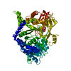
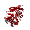
- PDB-5mst: Structure of the A domain of carboxylic acid reductase (CAR) from... -
+ Open data

- Basic information
| Entry | Database: PDB / ID: 5mst | |||||||||
|---|---|---|---|---|---|---|---|---|---|---|
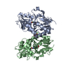
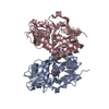
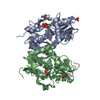
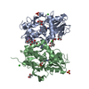
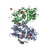
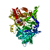
| Title | Structure of the A domain of carboxylic acid reductase (CAR) from Segniliparus rugosus in complex with AMP and a co-purified carboxylic acid | |||||||||
 Components Components | Thioester reductase domain-containing protein | |||||||||
 Keywords Keywords | OXIDOREDUCTASE / adenylation domain / carboxylic acid reductase | |||||||||
| Function / homology |  Function and homology information Function and homology informationlong-chain fatty acid-CoA ligase activity / Oxidoreductases; Acting on the aldehyde or oxo group of donors; With NAD+ or NADP+ as acceptor / oxidoreductase activity, acting on the aldehyde or oxo group of donors, NAD or NADP as acceptor / phosphopantetheine binding / NADP binding / ATP binding / membrane Similarity search - Function | |||||||||
| Biological species |  Segniliparus rugosus ATCC BAA-974 (bacteria) Segniliparus rugosus ATCC BAA-974 (bacteria) | |||||||||
| Method |  X-RAY DIFFRACTION / SYNCHROTRON / MOLECULAR REPLACEMENT / Resolution: 1.72 Å X-RAY DIFFRACTION / SYNCHROTRON / MOLECULAR REPLACEMENT / Resolution: 1.72 Å | |||||||||
 Authors Authors | Gahloth, D. / Leys, D. | |||||||||
 Citation Citation | Journal: Nat. Chem. Biol. / Year: 2017 Title: Structures of carboxylic acid reductase reveal domain dynamics underlying catalysis. Authors: Gahloth, D. / Dunstan, M.S. / Quaglia, D. / Klumbys, E. / Lockhart-Cairns, M.P. / Hill, A.M. / Derrington, S.R. / Scrutton, N.S. / Turner, N.J. / Leys, D. | |||||||||
| History |
|
- Structure visualization
| Structure viewer | Molecule: MolmilJmol/JSmol |
|---|
- Downloads & links
-Download
| PDBx/mmCIF format | 5mst.cif.gz | 317.4 KB | Display | PDBx/mmCIF format |
|---|---|---|---|---|
| PDB format | pdb5mst.ent.gz | 235.5 KB | Display | PDB format |
| PDBx/mmJSON format | 5mst.json.gz | Tree view | PDBx/mmJSON format | |
| Others |  Other downloads Other downloads |
-Validation report
| Arichive directory | https://data.pdbj.org/pub/pdb/validation_reports/ms/5mstftp://data.pdbj.org/pub/pdb/validation_reports/ms/5mst | HTTPS FTP |
|---|
-Related structure data
| Related structure data |  5mscC  5msdC  5msoC 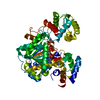 5mspC  5msrC  5mssC  5msuC  5msvC  5mswC  5msqS S: Starting model for refinement C: citing same article ( |
|---|---|
| Similar structure data |

-Links
 PDBj
PDBj


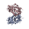
- Assembly
| Deposited unit | 
| ||||||||
|---|---|---|---|---|---|---|---|---|---|
| 1 | 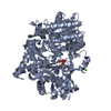
| ||||||||
| 2 | 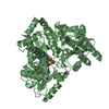
| ||||||||
| Unit cell |
|
-Components
| #1: Protein | Mass: 128368.164 Da / Num. of mol.: 2 Source method: isolated from a genetically manipulated source Source: (gene. exp.) Segniliparus rugosus ATCC BAA-974 (bacteria)Gene: HMPREF9336_01297 / Production host: #2: Chemical | ChemComp-CA /   Mass: 40.078 Da / Num. of mol.: 4 / Source method: obtained synthetically / Formula: Ca Mass: 40.078 Da / Num. of mol.: 4 / Source method: obtained synthetically / Formula: Ca#3: Chemical | 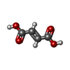  Mass: 116.072 Da / Num. of mol.: 2 / Source method: obtained synthetically / Formula: C4H4O4 Mass: 116.072 Da / Num. of mol.: 2 / Source method: obtained synthetically / Formula: C4H4O4#4: Chemical |   Mass: 347.221 Da / Num. of mol.: 2 / Source method: obtained synthetically / Formula: C10H14N5O7P / Comment: AMP*YM Mass: 347.221 Da / Num. of mol.: 2 / Source method: obtained synthetically / Formula: C10H14N5O7P / Comment: AMP*YM#5: Water | ChemComp-HOH / |  Mass: 18.015 Da / Num. of mol.: 1680 / Source method: isolated from a natural source / Formula: H2O Mass: 18.015 Da / Num. of mol.: 1680 / Source method: isolated from a natural source / Formula: H2O |
|---|
-Experimental details
-Experiment
| Experiment | Method: X-RAY DIFFRACTION / Number of used crystals: 1 |
|---|
- Sample preparation
| Crystal | Density Matthews: 1.82 Å3/Da / Density % sol: 32.47 % |
|---|---|
| Crystal grow | Temperature: 277 K / Method: vapor diffusion, sitting drop Details: Crystals of CARsr A domain were obtained in 0.2 M calcium chloride hydrate and 20% PEG3350. |
-Data collection
| Diffraction | Mean temperature: 100 K |
|---|---|
| Diffraction source | Source: SYNCHROTRON / Site: Diamond  / Beamline: I03 / Wavelength: 0.9763 Å / Beamline: I03 / Wavelength: 0.9763 Å |
| Detector | Type: DECTRIS PILATUS 6M-F / Detector: PIXEL / Date: May 15, 2016 |
| Radiation | Protocol: SINGLE WAVELENGTH / Monochromatic (M) / Laue (L): M / Scattering type: x-ray |
| Radiation wavelength | Wavelength: 0.9763 Å / Relative weight: 1 |
| Reflection | Resolution: 1.72→113.64 Å / Num. obs: 147465 / % possible obs: 99 % / Redundancy: 3.3 % / CC1/2: 1 / Rmerge(I) obs: 0.08 / Net I/σ(I): 12.8 |
| Reflection shell | Resolution: 1.72→1.78 Å / CC1/2: 0.4 / % possible all: 99.5 |
- Processing
| Software |
| ||||||||||||||||||||||||
|---|---|---|---|---|---|---|---|---|---|---|---|---|---|---|---|---|---|---|---|---|---|---|---|---|---|
| Refinement | Method to determine structure: MOLECULAR REPLACEMENT Starting model: 5MSQ Resolution: 1.72→113.64 Å / SU ML: 0.21 / Cross valid method: FREE R-VALUE / σ(F): 1.34 / Phase error: 22.06
| ||||||||||||||||||||||||
| Solvent computation | Shrinkage radii: 0.9 Å / VDW probe radii: 1.11 Å | ||||||||||||||||||||||||
| Refinement step | Cycle: LAST / Resolution: 1.72→113.64 Å
| ||||||||||||||||||||||||
| Refine LS restraints |
|
