 Movie
Movie Controller


+ Open data

- Basic information
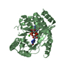
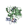
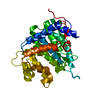
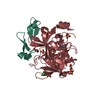
| Entry | Database: PDB / ID: 5mfu | |||||||||
|---|---|---|---|---|---|---|---|---|---|---|
| Title | PA3825-EAL Mn-pGpG Structure | |||||||||
 Components Components |
| |||||||||
 Keywords Keywords | HYDROLASE / EAL / Phosphodiesterase / biofilm formation / P Aeruginosa / PA3825 | |||||||||
| Function / homology |  Function and homology information Function and homology informationcyclic-guanylate-specific phosphodiesterase / regulation of single-species biofilm formation / cyclic-guanylate-specific phosphodiesterase activity / nucleotide binding / metal ion binding / plasma membrane Similarity search - Function | |||||||||
| Biological species |   Pseudomonas aeruginosa (bacteria) Pseudomonas aeruginosa (bacteria)synthetic construct (others) | |||||||||
| Method |  X-RAY DIFFRACTION / SYNCHROTRON / MOLECULAR REPLACEMENT / Resolution: 2.15 Å X-RAY DIFFRACTION / SYNCHROTRON / MOLECULAR REPLACEMENT / Resolution: 2.15 Å | |||||||||
 Authors Authors | Horrell, S. / Bellini, D. / Strange, R. / Wagner, A. / Walsh, M. | |||||||||
 Citation Citation | Journal: Sci Rep / Year: 2017 Title: Dimerisation induced formation of the active site and the identification of three metal sites in EAL-phosphodiesterases. Authors: Bellini, D. / Horrell, S. / Hutchin, A. / Phippen, C.W. / Strange, R.W. / Cai, Y. / Wagner, A. / Webb, J.S. / Tews, I. / Walsh, M.A. | |||||||||
| History |
|
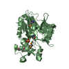
- Structure visualization
| Structure viewer | Molecule: MolmilJmol/JSmol |
|---|
- Downloads & links
-Download
| PDBx/mmCIF format | 5mfu.cif.gz | 71.5 KB | Display | PDBx/mmCIF format |
|---|---|---|---|---|
| PDB format | pdb5mfu.ent.gz | 50.6 KB | Display | PDB format |
| PDBx/mmJSON format | 5mfu.json.gz | Tree view | PDBx/mmJSON format | |
| Others |  Other downloads Other downloads |
-Validation report
| Arichive directory | https://data.pdbj.org/pub/pdb/validation_reports/mf/5mfuftp://data.pdbj.org/pub/pdb/validation_reports/mf/5mfu | HTTPS FTP |
|---|
-Related structure data
| Related structure data |  4y9mSC  5m1tC  5mf5C  5mkgC S: Starting model for refinement C: citing same article ( |
|---|---|
| Similar structure data |


-Links
 PDBj
PDBj- Assembly
| Deposited unit | 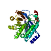
| ||||||||
|---|---|---|---|---|---|---|---|---|---|
| 1 |
| ||||||||
| Unit cell |
|
-Components
| #1: Protein | Mass: 28225.248 Da / Num. of mol.: 1 Source method: isolated from a genetically manipulated source Source: (gene. exp.) Pseudomonas aeruginosa (bacteria) / Gene: PA3825 / Production host: References: UniProt: Q9HXH7, cyclic-guanylate-specific phosphodiesterase |
|---|---|
| #2: RNA chain | Mass: 645.454 Da / Num. of mol.: 1 / Source method: obtained synthetically / Source: (synth.) synthetic construct (others) |
| #3: Chemical | ChemComp-MN /   Mass: 54.938 Da / Num. of mol.: 1 / Source method: obtained synthetically / Formula: Mn Mass: 54.938 Da / Num. of mol.: 1 / Source method: obtained synthetically / Formula: Mn |
| #4: Chemical | ChemComp-NA /   Mass: 22.990 Da / Num. of mol.: 1 / Source method: obtained synthetically / Formula: Na Mass: 22.990 Da / Num. of mol.: 1 / Source method: obtained synthetically / Formula: Na |
| #5: Water | ChemComp-HOH /  Mass: 18.015 Da / Num. of mol.: 118 / Source method: isolated from a natural source / Formula: H2O Mass: 18.015 Da / Num. of mol.: 118 / Source method: isolated from a natural source / Formula: H2O |
| Has protein modification | N |
-Experimental details
-Experiment
| Experiment | Method: X-RAY DIFFRACTION / Number of used crystals: 1 |
|---|
- Sample preparation
| Crystal | Density Matthews: 2.52 Å3/Da / Density % sol: 51.26 % |
|---|---|
| Crystal grow | Temperature: 277 K / Method: vapor diffusion, sitting drop / pH: 5.2 Details: 6-11% isopropanol, 0.1 M MES pH 6.5, 0.1 M sodium acetate pH 4.5 and 0.2 M MnCl2 |
-Data collection
| Diffraction | Mean temperature: 100 K |
|---|---|
| Diffraction source | Source: SYNCHROTRON / Site: Diamond  / Beamline: I02 / Wavelength: 1.8785 Å / Beamline: I02 / Wavelength: 1.8785 Å |
| Detector | Type: DECTRIS PILATUS 6M / Detector: PIXEL / Date: Aug 7, 2014 |
| Radiation | Protocol: SINGLE WAVELENGTH / Monochromatic (M) / Laue (L): M / Scattering type: x-ray |
| Radiation wavelength | Wavelength: 1.8785 Å / Relative weight: 1 |
| Reflection | Resolution: 2.15→27.16 Å / Num. obs: 16288 / % possible obs: 98.8 % / Redundancy: 3.8 % / Net I/σ(I): 6.6 |
- Processing
| Software |
| |||||||||||||||||||||||||||||||||||||||||||||||||||||||||||||||||||||||||||
|---|---|---|---|---|---|---|---|---|---|---|---|---|---|---|---|---|---|---|---|---|---|---|---|---|---|---|---|---|---|---|---|---|---|---|---|---|---|---|---|---|---|---|---|---|---|---|---|---|---|---|---|---|---|---|---|---|---|---|---|---|---|---|---|---|---|---|---|---|---|---|---|---|---|---|---|---|
| Refinement | Method to determine structure: MOLECULAR REPLACEMENT Starting model: 4Y9M Resolution: 2.15→26.66 Å / Cor.coef. Fo:Fc: 0.944 / Cor.coef. Fo:Fc free: 0.913 / SU B: 6.728 / SU ML: 0.172 / Cross valid method: THROUGHOUT / σ(F): 0 / ESU R: 0.257 / ESU R Free: 0.217 / Stereochemistry target values: MAXIMUM LIKELIHOOD Details: HYDROGENS HAVE BEEN ADDED IN THE RIDING POSITIONS U VALUES : REFINED INDIVIDUALLY
| |||||||||||||||||||||||||||||||||||||||||||||||||||||||||||||||||||||||||||
| Solvent computation | Ion probe radii: 0.8 Å / Shrinkage radii: 0.8 Å / VDW probe radii: 1.2 Å / Solvent model: MASK | |||||||||||||||||||||||||||||||||||||||||||||||||||||||||||||||||||||||||||
| Displacement parameters | Biso max: 146.96 Å2 / Biso mean: 36.452 Å2 / Biso min: 13.34 Å2
| |||||||||||||||||||||||||||||||||||||||||||||||||||||||||||||||||||||||||||
| Refinement step | Cycle: final / Resolution: 2.15→26.66 Å
| |||||||||||||||||||||||||||||||||||||||||||||||||||||||||||||||||||||||||||
| Refine LS restraints |
| |||||||||||||||||||||||||||||||||||||||||||||||||||||||||||||||||||||||||||
| LS refinement shell | Resolution: 2.15→2.206 Å / Total num. of bins used: 20
|
