 Movie
Movie Controller


+ Open data

- Basic information
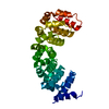
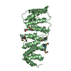
| Entry | Database: PDB / ID: 5mfn | ||||||
|---|---|---|---|---|---|---|---|
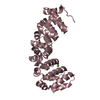
| Title | Designed armadillo repeat protein YIIIM5AII | ||||||
 Components Components | YIIIM5AII | ||||||
 Keywords Keywords | DE NOVO PROTEIN / Designed armadillo repeat protein / peptide binding | ||||||
| Function / homology | Leucine-rich Repeat Variant / Leucine-rich Repeat Variant / Alpha Horseshoe / Mainly Alpha / D-MALATE Function and homology information Function and homology information | ||||||
| Biological species | synthetic construct (others) | ||||||
| Method |  X-RAY DIFFRACTION / SYNCHROTRON / MOLECULAR REPLACEMENT / Resolution: 2.8 Å X-RAY DIFFRACTION / SYNCHROTRON / MOLECULAR REPLACEMENT / Resolution: 2.8 Å | ||||||
 Authors Authors | Hansen, S. / Ernst, P. / Reichen, C. / Ewald, C. / Mittl, P. / Plueckthun, A. | ||||||
 Citation Citation | Journal: J. Struct. Biol. / Year: 2018 Title: Curvature of designed armadillo repeat proteins allows modular peptide binding. Authors: Hansen, S. / Ernst, P. / Konig, S.L.B. / Reichen, C. / Ewald, C. / Nettels, D. / Mittl, P.R.E. / Schuler, B. / Pluckthun, A. | ||||||
| History |
|
- Structure visualization
| Structure viewer | Molecule: MolmilJmol/JSmol |
|---|
- Downloads & links
-Download
| PDBx/mmCIF format | 5mfn.cif.gz | 221.5 KB | Display | PDBx/mmCIF format |
|---|---|---|---|---|
| PDB format | pdb5mfn.ent.gz | 183.1 KB | Display | PDB format |
| PDBx/mmJSON format | 5mfn.json.gz | Tree view | PDBx/mmJSON format | |
| Others |  Other downloads Other downloads |
-Validation report
| Arichive directory | https://data.pdbj.org/pub/pdb/validation_reports/mf/5mfnftp://data.pdbj.org/pub/pdb/validation_reports/mf/5mfn | HTTPS FTP |
|---|
-Related structure data
| Related structure data |  5mfbC  5mfeC  5mffC  5mfgC  5mfhC  5mfiC  5mfjC  5mfkC  5mflC  5mfmC  5mfoC  5mfcS S: Starting model for refinement C: citing same article ( |
|---|---|
| Similar structure data |


-Links
 PDBj
PDBj


- Assembly
| Deposited unit | 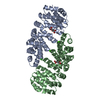
| ||||||||
|---|---|---|---|---|---|---|---|---|---|
| 1 | 
| ||||||||
| 2 | 
| ||||||||
| Unit cell |
|
-Components
| #1: Protein | Mass: 30027.445 Da / Num. of mol.: 2 Source method: isolated from a genetically manipulated source Source: (gene. exp.) synthetic construct (others) / Production host:  #2: Chemical |   Mass: 40.078 Da / Num. of mol.: 3 / Source method: obtained synthetically / Formula: Ca Mass: 40.078 Da / Num. of mol.: 3 / Source method: obtained synthetically / Formula: Ca#3: Chemical |   Mass: 134.087 Da / Num. of mol.: 2 / Source method: obtained synthetically / Formula: C4H6O5 Mass: 134.087 Da / Num. of mol.: 2 / Source method: obtained synthetically / Formula: C4H6O5#4: Water | ChemComp-HOH / |  Mass: 18.015 Da / Num. of mol.: 16 / Source method: isolated from a natural source / Formula: H2O Mass: 18.015 Da / Num. of mol.: 16 / Source method: isolated from a natural source / Formula: H2O |
|---|
-Experimental details
-Experiment
| Experiment | Method: X-RAY DIFFRACTION / Number of used crystals: 1 |
|---|
- Sample preparation
| Crystal | Density Matthews: 3.47 Å3/Da / Density % sol: 64.56 % |
|---|---|
| Crystal grow | Temperature: 277 K / Method: vapor diffusion, sitting drop / pH: 7 / Details: Sodium malonate 2.4M pH 7.0, 0.2M CaCl2 |
-Data collection
| Diffraction | Mean temperature: 100 K |
|---|---|
| Diffraction source | Source: SYNCHROTRON / Site: SLS  / Beamline: X06SA / Wavelength: 1 Å / Beamline: X06SA / Wavelength: 1 Å |
| Detector | Type: DECTRIS PILATUS 6M / Detector: PIXEL / Date: Feb 7, 2014 |
| Radiation | Protocol: SINGLE WAVELENGTH / Monochromatic (M) / Laue (L): M / Scattering type: x-ray |
| Radiation wavelength | Wavelength: 1 Å / Relative weight: 1 |
| Reflection | Resolution: 2.8→46.66 Å / Num. obs: 21564 / % possible obs: 99.9 % / Redundancy: 39.7 % / Biso Wilson estimate: 89.3 Å2 / CC1/2: 1 / Rmerge(I) obs: 0.257 / Net I/σ(I): 18.68 |
| Reflection shell | Resolution: 2.8→2.87 Å / Redundancy: 37.4 % / Rmerge(I) obs: 7.643 / Mean I/σ(I) obs: 0.69 / CC1/2: 0.184 / % possible all: 99.9 |
- Processing
| Software |
| ||||||||||||||||||||||||||||||||||||||||||||||||||||||||||||||||||||||||||||||||||||||||||||||||||||||||||||||||||
|---|---|---|---|---|---|---|---|---|---|---|---|---|---|---|---|---|---|---|---|---|---|---|---|---|---|---|---|---|---|---|---|---|---|---|---|---|---|---|---|---|---|---|---|---|---|---|---|---|---|---|---|---|---|---|---|---|---|---|---|---|---|---|---|---|---|---|---|---|---|---|---|---|---|---|---|---|---|---|---|---|---|---|---|---|---|---|---|---|---|---|---|---|---|---|---|---|---|---|---|---|---|---|---|---|---|---|---|---|---|---|---|---|---|---|---|
| Refinement | Method to determine structure: MOLECULAR REPLACEMENT Starting model: 5MFC Resolution: 2.8→46.66 Å / Cor.coef. Fo:Fc: 0.943 / Cor.coef. Fo:Fc free: 0.92 / SU R Cruickshank DPI: 0.517 / Cross valid method: THROUGHOUT / σ(F): 0 / SU R Blow DPI: 0.523 / SU Rfree Blow DPI: 0.299 / SU Rfree Cruickshank DPI: 0.303
| ||||||||||||||||||||||||||||||||||||||||||||||||||||||||||||||||||||||||||||||||||||||||||||||||||||||||||||||||||
| Displacement parameters | Biso mean: 131.96 Å2
| ||||||||||||||||||||||||||||||||||||||||||||||||||||||||||||||||||||||||||||||||||||||||||||||||||||||||||||||||||
| Refine analyze | Luzzati coordinate error obs: 0.51 Å | ||||||||||||||||||||||||||||||||||||||||||||||||||||||||||||||||||||||||||||||||||||||||||||||||||||||||||||||||||
| Refinement step | Cycle: 1 / Resolution: 2.8→46.66 Å
| ||||||||||||||||||||||||||||||||||||||||||||||||||||||||||||||||||||||||||||||||||||||||||||||||||||||||||||||||||
| Refine LS restraints |
| ||||||||||||||||||||||||||||||||||||||||||||||||||||||||||||||||||||||||||||||||||||||||||||||||||||||||||||||||||
| LS refinement shell | Resolution: 2.8→2.94 Å / Rfactor Rfree error: 0 / Total num. of bins used: 11
| ||||||||||||||||||||||||||||||||||||||||||||||||||||||||||||||||||||||||||||||||||||||||||||||||||||||||||||||||||
| Refinement TLS params. | Method: refined / Refine-ID: X-RAY DIFFRACTION
| ||||||||||||||||||||||||||||||||||||||||||||||||||||||||||||||||||||||||||||||||||||||||||||||||||||||||||||||||||
| Refinement TLS group |
|
