 Movie
Movie Controller

[English] 日本語
 Yorodumi
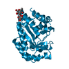
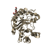
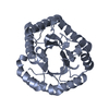
- PDB-5m42: Structure of Thermus thermophilus L-proline dehydrogenase lacking... -
+ Open data

- Basic information
| Entry | Database: PDB / ID: 5m42 | |||||||||
|---|---|---|---|---|---|---|---|---|---|---|
| Title | Structure of Thermus thermophilus L-proline dehydrogenase lacking alpha helices A, B and C | |||||||||
 Components Components | Proline dehydrogenase | |||||||||
 Keywords Keywords | OXIDOREDUCTASE / BETA8-ALPHA8-BARREL / FLAVOENZYME | |||||||||
| Function / homology |  Function and homology information Function and homology informationL-proline catabolic process / proline dehydrogenase / proline dehydrogenase activity / : / FAD binding / protein homodimerization activity Similarity search - Function | |||||||||
| Biological species |   Thermus thermophilus HB27 (bacteria) Thermus thermophilus HB27 (bacteria) | |||||||||
| Method |  X-RAY DIFFRACTION / SYNCHROTRON / MOLECULAR REPLACEMENT / Resolution: 2.2 Å X-RAY DIFFRACTION / SYNCHROTRON / MOLECULAR REPLACEMENT / Resolution: 2.2 Å | |||||||||
 Authors Authors | Martinez-Julvez, M. / Huijbers, M.M.E. / van Berkel, W.J.H. / Medina, M. | |||||||||
| Funding support |  Spain, Spain,  Netherlands, 2items Netherlands, 2items
| |||||||||
 Citation Citation | Journal: Sci Rep / Year: 2017 Title: Proline dehydrogenase from Thermus thermophilus does not discriminate between FAD and FMN as cofactor. Authors: Huijbers, M.M. / Martinez-Julvez, M. / Westphal, A.H. / Delgado-Arciniega, E. / Medina, M. / van Berkel, W.J. | |||||||||
| History |
|
- Structure visualization
| Structure viewer | Molecule: MolmilJmol/JSmol |
|---|
- Downloads & links
-Download
| PDBx/mmCIF format | 5m42.cif.gz | 66.7 KB | Display | PDBx/mmCIF format |
|---|---|---|---|---|
| PDB format | pdb5m42.ent.gz | 47.5 KB | Display | PDB format |
| PDBx/mmJSON format | 5m42.json.gz | Tree view | PDBx/mmJSON format | |
| Others |  Other downloads Other downloads |
-Validation report
| Arichive directory | https://data.pdbj.org/pub/pdb/validation_reports/m4/5m42ftp://data.pdbj.org/pub/pdb/validation_reports/m4/5m42 | HTTPS FTP |
|---|
-Related structure data
| Related structure data |  2g37S S: Starting model for refinement |
|---|---|
| Similar structure data |


-Links
 PDBj
PDBj- Assembly
| Deposited unit | 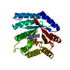
| ||||||||
|---|---|---|---|---|---|---|---|---|---|
| 1 |
| ||||||||
| Unit cell |
|
-Components
| #1: Protein | Mass: 32426.514 Da / Num. of mol.: 1 / Mutation: Delection of alpha helices A,B and C Source method: isolated from a genetically manipulated source Source: (gene. exp.) Thermus thermophilus HB27 (bacteria) / Gene: TT_C1214 / Production host: |
|---|---|
| #2: Chemical | ChemComp-FMN /   Mass: 456.344 Da / Num. of mol.: 1 / Source method: obtained synthetically / Formula: C17H21N4O9P Mass: 456.344 Da / Num. of mol.: 1 / Source method: obtained synthetically / Formula: C17H21N4O9P |
| #3: Water | ChemComp-HOH /  Mass: 18.015 Da / Num. of mol.: 64 / Source method: isolated from a natural source / Formula: H2O Mass: 18.015 Da / Num. of mol.: 64 / Source method: isolated from a natural source / Formula: H2O |
-Experimental details
-Experiment
| Experiment | Method: X-RAY DIFFRACTION / Number of used crystals: 1 |
|---|
- Sample preparation
| Crystal | Density Matthews: 2.84 Å3/Da / Density % sol: 56.7 % |
|---|---|
| Crystal grow | Temperature: 291 K / Method: vapor diffusion, sitting drop / pH: 4.6 Details: 20 % v/v 2-propanol, 0.2 M calcium chloride dihydrate and 0.1 M sodium acetate pH 4.6 |
-Data collection
| Diffraction | Mean temperature: 100 K |
|---|---|
| Diffraction source | Source: SYNCHROTRON / Site: Diamond  / Beamline: I02 / Wavelength: 0.97949 Å / Beamline: I02 / Wavelength: 0.97949 Å |
| Detector | Type: DECTRIS PILATUS 6M-F / Detector: PIXEL / Date: May 14, 2015 |
| Radiation | Protocol: SINGLE WAVELENGTH / Monochromatic (M) / Laue (L): M / Scattering type: x-ray |
| Radiation wavelength | Wavelength: 0.97949 Å / Relative weight: 1 |
| Reflection | Resolution: 2.2→65.96 Å / Num. obs: 18892 / % possible obs: 100 % / Redundancy: 8.4 % / Rmerge(I) obs: 0.139 / Net I/σ(I): 10.9 |
| Reflection shell | Resolution: 2.2→2.32 Å / Redundancy: 8.7 % / % possible all: 100 |
- Processing
| Software |
| ||||||||||||||||||||||||||||||||||||||||||||||||||||||||||||||||||||||||||||||||||||||||||||||||||||||||||||||||||||||||||||||||||||||||||||||||||||||||||||||||||||||||||||||||||||||
|---|---|---|---|---|---|---|---|---|---|---|---|---|---|---|---|---|---|---|---|---|---|---|---|---|---|---|---|---|---|---|---|---|---|---|---|---|---|---|---|---|---|---|---|---|---|---|---|---|---|---|---|---|---|---|---|---|---|---|---|---|---|---|---|---|---|---|---|---|---|---|---|---|---|---|---|---|---|---|---|---|---|---|---|---|---|---|---|---|---|---|---|---|---|---|---|---|---|---|---|---|---|---|---|---|---|---|---|---|---|---|---|---|---|---|---|---|---|---|---|---|---|---|---|---|---|---|---|---|---|---|---|---|---|---|---|---|---|---|---|---|---|---|---|---|---|---|---|---|---|---|---|---|---|---|---|---|---|---|---|---|---|---|---|---|---|---|---|---|---|---|---|---|---|---|---|---|---|---|---|---|---|---|---|
| Refinement | Method to determine structure: MOLECULAR REPLACEMENT Starting model: 2G37 Resolution: 2.2→60.01 Å / Cor.coef. Fo:Fc: 0.958 / Cor.coef. Fo:Fc free: 0.94 / SU B: 5.356 / SU ML: 0.13 / Cross valid method: THROUGHOUT / ESU R: 0.189 / ESU R Free: 0.171
| ||||||||||||||||||||||||||||||||||||||||||||||||||||||||||||||||||||||||||||||||||||||||||||||||||||||||||||||||||||||||||||||||||||||||||||||||||||||||||||||||||||||||||||||||||||||
| Solvent computation | Ion probe radii: 0.8 Å / Shrinkage radii: 0.8 Å / VDW probe radii: 1.2 Å | ||||||||||||||||||||||||||||||||||||||||||||||||||||||||||||||||||||||||||||||||||||||||||||||||||||||||||||||||||||||||||||||||||||||||||||||||||||||||||||||||||||||||||||||||||||||
| Displacement parameters | Biso mean: 37.672 Å2
| ||||||||||||||||||||||||||||||||||||||||||||||||||||||||||||||||||||||||||||||||||||||||||||||||||||||||||||||||||||||||||||||||||||||||||||||||||||||||||||||||||||||||||||||||||||||
| Refinement step | Cycle: 1 / Resolution: 2.2→60.01 Å
| ||||||||||||||||||||||||||||||||||||||||||||||||||||||||||||||||||||||||||||||||||||||||||||||||||||||||||||||||||||||||||||||||||||||||||||||||||||||||||||||||||||||||||||||||||||||
| Refine LS restraints |
|