 Movie
Movie Controller

[English] 日本語
 Yorodumi
- PDB-5m2q: Structure of cobinamide-bound BtuF mutant W66F, the periplasmic v... -
+ Open data

- Basic information
| Entry | Database: PDB / ID: 5m2q | ||||||
|---|---|---|---|---|---|---|---|
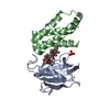
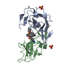
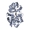
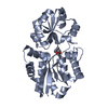
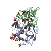
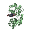
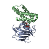
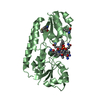
| Title | Structure of cobinamide-bound BtuF mutant W66F, the periplasmic vitamin B12 binding protein in E.coli | ||||||
 Components Components | Outer membrane protein A,Vitamin B12-binding protein | ||||||
 Keywords Keywords | TRANSPORT PROTEIN / BtuF / Cobinamide / periplasmic binding protein / ABC transporter | ||||||
| Function / homology |  Function and homology information Function and homology informationcobalamin transport complex / outer membrane protein complex / cobalamin transport / monoatomic ion transmembrane transporter activity / detection of virus / cobalamin binding / outer membrane / porin activity / pore complex / monoatomic ion transport ...cobalamin transport complex / outer membrane protein complex / cobalamin transport / monoatomic ion transmembrane transporter activity / detection of virus / cobalamin binding / outer membrane / porin activity / pore complex / monoatomic ion transport / cell outer membrane / outer membrane-bounded periplasmic space / periplasmic space / symbiont entry into host cell / DNA damage response / membrane / identical protein binding Similarity search - Function | ||||||
| Biological species |  | ||||||
| Method |  X-RAY DIFFRACTION / SYNCHROTRON / MOLECULAR REPLACEMENT / Resolution: 1.7 Å X-RAY DIFFRACTION / SYNCHROTRON / MOLECULAR REPLACEMENT / Resolution: 1.7 Å | ||||||
 Authors Authors | Mireku, S.A. / Ruetz, M. / Zhou, T. / Korkhov, V.M. / Kraeutler, B. / Locher, K.P. | ||||||
 Citation Citation | Journal: Sci Rep / Year: 2017 Title: Conformational Change of a Tryptophan Residue in BtuF Facilitates Binding and Transport of Cobinamide by the Vitamin B12 Transporter BtuCD-F. Authors: Mireku, S.A. / Ruetz, M. / Zhou, T. / Korkhov, V.M. / Krautler, B. / Locher, K.P. | ||||||
| History |
|
- Structure visualization
| Structure viewer | Molecule: MolmilJmol/JSmol |
|---|
- Downloads & links
-Download
| PDBx/mmCIF format | 5m2q.cif.gz | 207 KB | Display | PDBx/mmCIF format |
|---|---|---|---|---|
| PDB format | pdb5m2q.ent.gz | 163.8 KB | Display | PDB format |
| PDBx/mmJSON format | 5m2q.json.gz | Tree view | PDBx/mmJSON format | |
| Others |  Other downloads Other downloads |
-Validation report
| Arichive directory | https://data.pdbj.org/pub/pdb/validation_reports/m2/5m2qftp://data.pdbj.org/pub/pdb/validation_reports/m2/5m2q | HTTPS FTP |
|---|
-Related structure data
| Related structure data |  5m29SC  5m34C  5m3bC S: Starting model for refinement C: citing same article ( |
|---|---|
| Similar structure data |


-Links
 PDBj
PDBj


- Assembly
| Deposited unit | 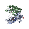
| ||||||||
|---|---|---|---|---|---|---|---|---|---|
| 1 | 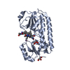
| ||||||||
| 2 | 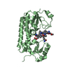
| ||||||||
| Unit cell |
|
-Components
| #1: Protein | Mass: 31472.889 Da / Num. of mol.: 2 / Mutation: W66F Source method: isolated from a genetically manipulated source Details: OmpA-NcoI-BtuF-BamHI-3C-BamHI-His6 / Source: (gene. exp.) Gene: ompA, con, tolG, tut, b0957, JW0940, btuF, yadT, b0158, JW0154 Production host: #2: Chemical |   Mass: 990.087 Da / Num. of mol.: 2 / Source method: obtained synthetically / Formula: C48H72CoN11O8 Mass: 990.087 Da / Num. of mol.: 2 / Source method: obtained synthetically / Formula: C48H72CoN11O8#3: Chemical |   Mass: 26.017 Da / Num. of mol.: 2 / Source method: obtained synthetically / Formula: CN Mass: 26.017 Da / Num. of mol.: 2 / Source method: obtained synthetically / Formula: CN#4: Chemical |   Mass: 92.094 Da / Num. of mol.: 3 / Source method: obtained synthetically / Formula: C3H8O3 Mass: 92.094 Da / Num. of mol.: 3 / Source method: obtained synthetically / Formula: C3H8O3#5: Water | ChemComp-HOH / |  Mass: 18.015 Da / Num. of mol.: 390 / Source method: isolated from a natural source / Formula: H2O Mass: 18.015 Da / Num. of mol.: 390 / Source method: isolated from a natural source / Formula: H2OHas protein modification | Y | |
|---|
-Experimental details
-Experiment
| Experiment | Method: X-RAY DIFFRACTION / Number of used crystals: 1 |
|---|
- Sample preparation
| Crystal | Density Matthews: 2.33 Å3/Da / Density % sol: 47.32 % |
|---|---|
| Crystal grow | Temperature: 293.15 K / Method: vapor diffusion, sitting drop / Details: PEG3350 HEPES pH 7 Tryptone |
-Data collection
| Diffraction | Mean temperature: 123 K |
|---|---|
| Diffraction source | Source: SYNCHROTRON / Site: SLS  / Beamline: X06SA / Wavelength: 0.97794 Å / Beamline: X06SA / Wavelength: 0.97794 Å |
| Detector | Type: DECTRIS PILATUS 6M-F / Detector: PIXEL / Date: Dec 13, 2014 |
| Radiation | Protocol: SINGLE WAVELENGTH / Monochromatic (M) / Laue (L): M / Scattering type: x-ray |
| Radiation wavelength | Wavelength: 0.97794 Å / Relative weight: 1 |
| Reflection | Resolution: 1.7→19.71 Å / Num. obs: 61170 / % possible obs: 96.27 % / Redundancy: 3.1 % / Net I/σ(I): 11.05 |
- Processing
| Software |
| ||||||||||||||||||||||||||||||||||||||||||||||||||||||||||||||||||||||||||||||||||||||||||||||||||||||||||||||||||||||||||||||||||||||||||||||||||||||||||||||||||||||||||||||||||||||
|---|---|---|---|---|---|---|---|---|---|---|---|---|---|---|---|---|---|---|---|---|---|---|---|---|---|---|---|---|---|---|---|---|---|---|---|---|---|---|---|---|---|---|---|---|---|---|---|---|---|---|---|---|---|---|---|---|---|---|---|---|---|---|---|---|---|---|---|---|---|---|---|---|---|---|---|---|---|---|---|---|---|---|---|---|---|---|---|---|---|---|---|---|---|---|---|---|---|---|---|---|---|---|---|---|---|---|---|---|---|---|---|---|---|---|---|---|---|---|---|---|---|---|---|---|---|---|---|---|---|---|---|---|---|---|---|---|---|---|---|---|---|---|---|---|---|---|---|---|---|---|---|---|---|---|---|---|---|---|---|---|---|---|---|---|---|---|---|---|---|---|---|---|---|---|---|---|---|---|---|---|---|---|---|
| Refinement | Method to determine structure: MOLECULAR REPLACEMENT Starting model: 5M29 Resolution: 1.7→19.71 Å / Cor.coef. Fo:Fc: 0.962 / Cor.coef. Fo:Fc free: 0.944 / SU B: 8.866 / SU ML: 0.132 / Cross valid method: THROUGHOUT / ESU R: 0.114 / ESU R Free: 0.116 / Details: HYDROGENS HAVE BEEN ADDED IN THE RIDING POSITIONS
| ||||||||||||||||||||||||||||||||||||||||||||||||||||||||||||||||||||||||||||||||||||||||||||||||||||||||||||||||||||||||||||||||||||||||||||||||||||||||||||||||||||||||||||||||||||||
| Solvent computation | Ion probe radii: 0.8 Å / Shrinkage radii: 0.8 Å / VDW probe radii: 1.2 Å | ||||||||||||||||||||||||||||||||||||||||||||||||||||||||||||||||||||||||||||||||||||||||||||||||||||||||||||||||||||||||||||||||||||||||||||||||||||||||||||||||||||||||||||||||||||||
| Displacement parameters | Biso mean: 33.948 Å2
| ||||||||||||||||||||||||||||||||||||||||||||||||||||||||||||||||||||||||||||||||||||||||||||||||||||||||||||||||||||||||||||||||||||||||||||||||||||||||||||||||||||||||||||||||||||||
| Refinement step | Cycle: 1 / Resolution: 1.7→19.71 Å
| ||||||||||||||||||||||||||||||||||||||||||||||||||||||||||||||||||||||||||||||||||||||||||||||||||||||||||||||||||||||||||||||||||||||||||||||||||||||||||||||||||||||||||||||||||||||
| Refine LS restraints |
|