 Movie
Movie Controller

[English] 日本語
 Yorodumi
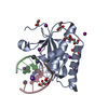
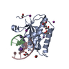
- PDB-5lz6: Crystal structure of human ACBD3 GOLD domain in complex with 3A p... -
+ Open data

- Basic information
| Entry | Database: PDB / ID: 5lz6 | ||||||
|---|---|---|---|---|---|---|---|
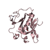
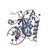
| Title | Crystal structure of human ACBD3 GOLD domain in complex with 3A protein of Aichivirus B | ||||||
 Components Components |
| ||||||
 Keywords Keywords | ANTIVIRAL PROTEIN / ACBD3 / GOLD / 3A / Aichivirus | ||||||
| Function / homology |  Function and homology information Function and homology informationfatty-acyl-CoA binding / steroid biosynthetic process / Golgi Associated Vesicle Biogenesis / protein kinase A regulatory subunit binding / cytoplasmic vesicle membrane / T=pseudo3 icosahedral viral capsid / host cell cytoplasmic vesicle membrane / channel activity / monoatomic ion transmembrane transport / RNA helicase activity ...fatty-acyl-CoA binding / steroid biosynthetic process / Golgi Associated Vesicle Biogenesis / protein kinase A regulatory subunit binding / cytoplasmic vesicle membrane / T=pseudo3 icosahedral viral capsid / host cell cytoplasmic vesicle membrane / channel activity / monoatomic ion transmembrane transport / RNA helicase activity / Golgi membrane / cysteine-type endopeptidase activity / viral RNA genome replication / RNA-directed RNA polymerase activity / symbiont entry into host cell / DNA-templated transcription / virion attachment to host cell / structural molecule activity / Golgi apparatus / mitochondrion / proteolysis / RNA binding / ATP binding / membrane Similarity search - Function | ||||||
| Biological species |  Homo sapiens (human) Homo sapiens (human) Aichivirus B Aichivirus B | ||||||
| Method |  X-RAY DIFFRACTION / SYNCHROTRON / MOLECULAR REPLACEMENT / Resolution: 2.6 Å X-RAY DIFFRACTION / SYNCHROTRON / MOLECULAR REPLACEMENT / Resolution: 2.6 Å | ||||||
 Authors Authors | Klima, M. / Boura, E. | ||||||
 Citation Citation | Journal: Structure / Year: 2017 Title: Kobuviral Non-structural 3A Proteins Act as Molecular Harnesses to Hijack the Host ACBD3 Protein. Authors: Klima, M. / Chalupska, D. / Rozycki, B. / Humpolickova, J. / Rezabkova, L. / Silhan, J. / Baumlova, A. / Dubankova, A. / Boura, E. | ||||||
| History |
|
- Structure visualization
| Structure viewer | Molecule: MolmilJmol/JSmol |
|---|
- Downloads & links
-Download
| PDBx/mmCIF format | 5lz6.cif.gz | 47 KB | Display | PDBx/mmCIF format |
|---|---|---|---|---|
| PDB format | pdb5lz6.ent.gz | 31.5 KB | Display | PDB format |
| PDBx/mmJSON format | 5lz6.json.gz | Tree view | PDBx/mmJSON format | |
| Others |  Other downloads Other downloads |
-Validation report
| Arichive directory | https://data.pdbj.org/pub/pdb/validation_reports/lz/5lz6ftp://data.pdbj.org/pub/pdb/validation_reports/lz/5lz6 | HTTPS FTP |
|---|
-Related structure data
| Related structure data | 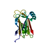 5lz1SC 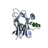 5lz3C S: Starting model for refinement C: citing same article ( |
|---|---|
| Similar structure data |


-Links
 PDBj
PDBj


- Assembly
| Deposited unit | 
| ||||||||
|---|---|---|---|---|---|---|---|---|---|
| 1 |
| ||||||||
| Unit cell |
|
-Components
| #1: Protein | Mass: 19261.217 Da / Num. of mol.: 1 Source method: isolated from a genetically manipulated source Source: (gene. exp.) Homo sapiens (human) / Gene: ACBD3, GCP60, GOCAP1, GOLPH1 / Production host:  |
|---|---|
| #2: Protein/peptide | Mass: 4182.599 Da / Num. of mol.: 1 Source method: isolated from a genetically manipulated source Source: (gene. exp.) Aichivirus B / Production host: |
| #3: Sugar | ChemComp-BGC /   Type: D-saccharide, beta linking / Mass: 180.156 Da / Num. of mol.: 1 Type: D-saccharide, beta linking / Mass: 180.156 Da / Num. of mol.: 1Source method: isolated from a genetically manipulated source Formula: C6H12O6 |
-Experimental details
-Experiment
| Experiment | Method: X-RAY DIFFRACTION / Number of used crystals: 1 |
|---|
- Sample preparation
| Crystal | Density Matthews: 2.75 Å3/Da / Density % sol: 55.25 % |
|---|---|
| Crystal grow | Temperature: 291 K / Method: vapor diffusion, sitting drop Details: 100mM MOPSO/Bis-Tris pH 6.5, 15% w/v PEG 3.000, 1% w/v DDAO, 10% 1,4-butanediol, 10% w/v glucose, 20mM L-arginine/L-threonine/L-histidine/betaine, 10mM trans-4-hydroxy-L-proline |
-Data collection
| Diffraction | Mean temperature: 100 K |
|---|---|
| Diffraction source | Source: SYNCHROTRON / Site: BESSY  / Beamline: 14.1 / Wavelength: 0.97981 Å / Beamline: 14.1 / Wavelength: 0.97981 Å |
| Detector | Type: DECTRIS PILATUS 6M / Detector: PIXEL / Date: Mar 17, 2016 |
| Radiation | Protocol: SINGLE WAVELENGTH / Monochromatic (M) / Laue (L): M / Scattering type: x-ray |
| Radiation wavelength | Wavelength: 0.97981 Å / Relative weight: 1 |
| Reflection | Resolution: 2.6→48.18 Å / Num. obs: 8472 / % possible obs: 99.96 % / Redundancy: 10.4 % / Biso Wilson estimate: 77.41 Å2 / CC1/2: 1 / Rmerge(I) obs: 0.07815 / Rsym value: 0.07815 / Net I/σ(I): 22.47 |
| Reflection shell | Resolution: 2.6→2.693 Å / Redundancy: 10.8 % / Rmerge(I) obs: 1.984 / Mean I/σ(I) obs: 1.24 / CC1/2: 0.454 / % possible all: 100 |
- Processing
| Software |
| ||||||||||||||||||||||||||||
|---|---|---|---|---|---|---|---|---|---|---|---|---|---|---|---|---|---|---|---|---|---|---|---|---|---|---|---|---|---|
| Refinement | Method to determine structure: MOLECULAR REPLACEMENT Starting model: 5LZ1 Resolution: 2.6→48.177 Å / SU ML: 0.46 / Cross valid method: FREE R-VALUE / σ(F): 1.36 / Phase error: 31.31
| ||||||||||||||||||||||||||||
| Solvent computation | Shrinkage radii: 0.9 Å / VDW probe radii: 1.11 Å | ||||||||||||||||||||||||||||
| Refinement step | Cycle: LAST / Resolution: 2.6→48.177 Å
| ||||||||||||||||||||||||||||
| Refine LS restraints |
| ||||||||||||||||||||||||||||
| LS refinement shell |
|
