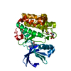
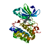
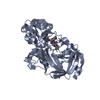
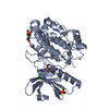
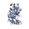
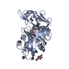
Entry Database : PDB / ID : 5lvlTitle Human PDK1 Kinase Domain in Complex with Compound PS653 Bound to the ATP-Binding Site 3-phosphoinositide-dependent protein kinase 1 Keywords / / / / Function / homology Function Domain/homology Component
/ / / / / / / / / / / / / / / / / / / / / / / / / / / / / / / / / / / / / / / / / / / / / / / / / / / / / / / / / / / / / / / / / / / / / / / / / / / / / / / / / / / / / / / / / / / / / / / / / / / / / / / Biological species Homo sapiens (human)Method / / / Resolution : 1.4 Å Authors Schulze, J.O. / Saladino, G. / Busschots, K. / Neimanis, S. / Suess, E. / Odadzic, D. / Zeuzem, S. / Hindie, V. / Herbrand, A.K. / Lisa, M.N. ...Schulze, J.O. / Saladino, G. / Busschots, K. / Neimanis, S. / Suess, E. / Odadzic, D. / Zeuzem, S. / Hindie, V. / Herbrand, A.K. / Lisa, M.N. / Alzari, P.M. / Gervasio, F.L. / Biondi, R.M. Funding support Organization Grant number Country BMBF GO-Bio 0315102 German Research Foundation BI 1044/2-3 German Research Foundation BI 1044/12-1
Journal : Cell Chem Biol / Year : 2016Title : Bidirectional Allosteric Communication between the ATP-Binding Site and the Regulatory PIF Pocket in PDK1 Protein Kinase.Authors : Schulze, J.O. / Saladino, G. / Busschots, K. / Neimanis, S. / Su, E. / Odadzic, D. / Zeuzem, S. / Hindie, V. / Herbrand, A.K. / Lisa, M.N. / Alzari, P.M. / Gervasio, F.L. / Biondi, R.M. History Deposition Sep 14, 2016 Deposition site / Processing site Revision 1.0 Oct 19, 2016 Provider / Type Revision 1.1 Nov 2, 2016 Group Revision 2.0 Oct 16, 2019 Group / Author supporting evidence / Data collectionCategory / pdbx_audit_support / reflns_shellItem / _pdbx_audit_support.funding_organizationRevision 2.1 Jan 17, 2024 Group / Database references / Refinement descriptionCategory chem_comp_atom / chem_comp_bond ... chem_comp_atom / chem_comp_bond / database_2 / pdbx_initial_refinement_model Item / _database_2.pdbx_database_accession
Show all Show less
 Movie
Movie Controller

 Yorodumi
 Open data
 Basic information
 Components
 Keywords
 Function and homology information
 Homo sapiens (human)
 X-RAY DIFFRACTION /
  Authors
 Germany, 3items
  Citation
 Structure visualization
 Downloads & links
 Other downloads
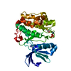


 PDBj
PDBj


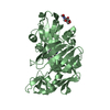
 Assembly

 Spodoptera frugiperda (fall armyworm)
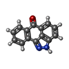
 Mass: 220.226 Da / Num. of mol.: 1 / Source method: obtained synthetically / Formula: C14H8N2O / Comment: inhibitor*YM
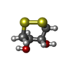
 Mass: 152.235 Da / Num. of mol.: 1 / Source method: obtained synthetically / Formula: C4H8O2S2
 Mass: 78.133 Da / Num. of mol.: 3 / Source method: obtained synthetically / Formula: C2H6OS / Comment: DMSO, precipitant*YM
 Mass: 18.015 Da / Num. of mol.: 267 / Source method: isolated from a natural source / Formula: H2O
 Sample preparation
 Processing