 Movie
Movie Controller

[English] 日本語
 Yorodumi
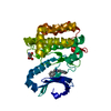
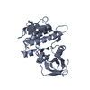
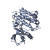
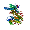
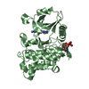
- PDB-5lvm: Human PDK1 Kinase Domain in Complex with Adenine Bound to the ATP... -
+ Open data

- Basic information
| Entry | Database: PDB / ID: 5lvm | ||||||||||||
|---|---|---|---|---|---|---|---|---|---|---|---|---|---|
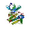
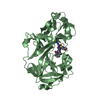
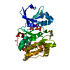
| Title | Human PDK1 Kinase Domain in Complex with Adenine Bound to the ATP-Binding Site | ||||||||||||
 Components Components | 3-phosphoinositide-dependent protein kinase 1 | ||||||||||||
 Keywords Keywords | TRANSFERASE / protein kinase / allosteric regulation / small compounds / PIF-pocket | ||||||||||||
| Function / homology |  Function and homology information Function and homology informationintracellular signaling cassette / 3-phosphoinositide-dependent protein kinase activity / Activation of AKT2 / regulation of mast cell degranulation / type B pancreatic cell development / negative regulation of toll-like receptor signaling pathway / RSK activation / positive regulation of vascular endothelial cell proliferation / regulation of canonical NF-kappaB signal transduction / phospholipase activator activity ...intracellular signaling cassette / 3-phosphoinositide-dependent protein kinase activity / Activation of AKT2 / regulation of mast cell degranulation / type B pancreatic cell development / negative regulation of toll-like receptor signaling pathway / RSK activation / positive regulation of vascular endothelial cell proliferation / regulation of canonical NF-kappaB signal transduction / phospholipase activator activity / positive regulation of sprouting angiogenesis / Constitutive Signaling by AKT1 E17K in Cancer / negative regulation of cardiac muscle cell apoptotic process / Role of LAT2/NTAL/LAB on calcium mobilization / CD28 dependent PI3K/Akt signaling / negative regulation of endothelial cell apoptotic process / positive regulation of blood vessel endothelial cell migration / SARS-CoV-2 targets host intracellular signalling and regulatory pathways / Estrogen-stimulated signaling through PRKCZ / extrinsic apoptotic signaling pathway / vascular endothelial cell response to laminar fluid shear stress / SARS-CoV-1 targets host intracellular signalling and regulatory pathways / RHO GTPases activate PKNs / GPVI-mediated activation cascade / phospholipase binding / T cell costimulation / Integrin signaling / cell projection / insulin-like growth factor receptor signaling pathway / cellular response to epidermal growth factor stimulus / positive regulation of release of sequestered calcium ion into cytosol / VEGFR2 mediated cell proliferation / VEGFR2 mediated vascular permeability / positive regulation of protein localization to plasma membrane / calcium-mediated signaling / phosphatidylinositol 3-kinase/protein kinase B signal transduction / negative regulation of transforming growth factor beta receptor signaling pathway / CLEC7A (Dectin-1) signaling / epidermal growth factor receptor signaling pathway / FCERI mediated NF-kB activation / cellular response to insulin stimulus / positive regulation of angiogenesis / insulin receptor signaling pathway / Regulation of TP53 Degradation / PIP3 activates AKT signaling / protein autophosphorylation / Downstream TCR signaling / G beta:gamma signalling through PI3Kgamma / cell migration / actin cytoskeleton organization / cytoplasmic vesicle / High laminar flow shear stress activates signaling by PIEZO1 and PECAM1:CDH5:KDR in endothelial cells / protein phosphorylation / positive regulation of phosphatidylinositol 3-kinase/protein kinase B signal transduction / non-specific serine/threonine protein kinase / postsynaptic density / intracellular signal transduction / protein serine kinase activity / focal adhesion / protein serine/threonine kinase activity / DNA-templated transcription / ATP binding / nucleus / plasma membrane / cytosol / cytoplasm Similarity search - Function | ||||||||||||
| Biological species |  Homo sapiens (human) Homo sapiens (human) | ||||||||||||
| Method |  X-RAY DIFFRACTION / SYNCHROTRON / MOLECULAR REPLACEMENT / Resolution: 1.26 Å X-RAY DIFFRACTION / SYNCHROTRON / MOLECULAR REPLACEMENT / Resolution: 1.26 Å | ||||||||||||
 Authors Authors | Schulze, J.O. / Saladino, G. / Busschots, K. / Neimanis, S. / Suess, E. / Odadzic, D. / Zeuzem, S. / Hindie, V. / Herbrand, A.K. / Lisa, M.N. ...Schulze, J.O. / Saladino, G. / Busschots, K. / Neimanis, S. / Suess, E. / Odadzic, D. / Zeuzem, S. / Hindie, V. / Herbrand, A.K. / Lisa, M.N. / Alzari, P.M. / Gervasio, F.L. / Biondi, R.M. | ||||||||||||
| Funding support |  Germany, 3items Germany, 3items
| ||||||||||||
 Citation Citation | Journal: Cell Chem Biol / Year: 2016 Title: Bidirectional Allosteric Communication between the ATP-Binding Site and the Regulatory PIF Pocket in PDK1 Protein Kinase. Authors: Schulze, J.O. / Saladino, G. / Busschots, K. / Neimanis, S. / Su, E. / Odadzic, D. / Zeuzem, S. / Hindie, V. / Herbrand, A.K. / Lisa, M.N. / Alzari, P.M. / Gervasio, F.L. / Biondi, R.M. | ||||||||||||
| History |
|
- Structure visualization
| Structure viewer | Molecule: MolmilJmol/JSmol |
|---|
- Downloads & links
-Download
| PDBx/mmCIF format | 5lvm.cif.gz | 191.5 KB | Display | PDBx/mmCIF format |
|---|---|---|---|---|
| PDB format | pdb5lvm.ent.gz | 154.6 KB | Display | PDB format |
| PDBx/mmJSON format | 5lvm.json.gz | Tree view | PDBx/mmJSON format | |
| Others |  Other downloads Other downloads |
-Validation report
| Arichive directory | https://data.pdbj.org/pub/pdb/validation_reports/lv/5lvmftp://data.pdbj.org/pub/pdb/validation_reports/lv/5lvm | HTTPS FTP |
|---|
-Related structure data
| Related structure data |  5lvlC 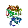 5lvnC  5lvoC  5lvpC  3hrcS S: Starting model for refinement C: citing same article ( |
|---|---|
| Similar structure data |


-Links
 PDBj
PDBj


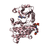
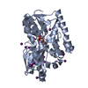
- Assembly
| Deposited unit | 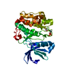
| ||||||||
|---|---|---|---|---|---|---|---|---|---|
| 1 |
| ||||||||
| Unit cell |
|
-Components
| #1: Protein | Mass: 35392.566 Da / Num. of mol.: 1 / Mutation: Y288G, Q292A Source method: isolated from a genetically manipulated source Source: (gene. exp.) Homo sapiens (human) / Gene: PDPK1, PDK1 / Production host:   Spodoptera frugiperda (fall armyworm) Spodoptera frugiperda (fall armyworm)References: UniProt: O15530, non-specific serine/threonine protein kinase |
|---|---|
| #2: Chemical | ChemComp-ADE /   Mass: 135.127 Da / Num. of mol.: 1 / Source method: obtained synthetically / Formula: C5H5N5 Mass: 135.127 Da / Num. of mol.: 1 / Source method: obtained synthetically / Formula: C5H5N5Details: Attention: "A" was not accepted as sample sequence. This is just adenine, not an RNA molecule. Source: (synth.) Homo sapiens (human) |
| #3: Chemical | ChemComp-DTD / 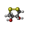  Mass: 152.235 Da / Num. of mol.: 1 / Source method: obtained synthetically / Formula: C4H8O2S2 Mass: 152.235 Da / Num. of mol.: 1 / Source method: obtained synthetically / Formula: C4H8O2S2 |
| #4: Water | ChemComp-HOH /  Mass: 18.015 Da / Num. of mol.: 302 / Source method: isolated from a natural source / Formula: H2O Mass: 18.015 Da / Num. of mol.: 302 / Source method: isolated from a natural source / Formula: H2O |
| Has protein modification | Y |
-Experimental details
-Experiment
| Experiment | Method: X-RAY DIFFRACTION / Number of used crystals: 1 |
|---|
- Sample preparation
| Crystal | Density Matthews: 2.13 Å3/Da / Density % sol: 42.35 % |
|---|---|
| Crystal grow | Temperature: 293 K / Method: vapor diffusion, hanging drop / pH: 7.5 / Details: 1.2 M NA CITRATE, 0.1 M HEPES PH 7.5, 10 MM DTT |
-Data collection
| Diffraction | Mean temperature: 100 K |
|---|---|
| Diffraction source | Source: SYNCHROTRON / Site: BESSY / Beamline: 14.1 / Wavelength: 0.91841 Å |
| Detector | Type: DECTRIS PILATUS 6M / Detector: PIXEL / Date: Jun 3, 2014 |
| Radiation | Protocol: SINGLE WAVELENGTH / Monochromatic (M) / Laue (L): M / Scattering type: x-ray |
| Radiation wavelength | Wavelength: 0.91841 Å / Relative weight: 1 |
| Reflection | Resolution: 1.26→44 Å / Num. obs: 82126 / % possible obs: 99.6 % / Redundancy: 6.6 % / Net I/σ(I): 17.8 |
- Processing
| Software |
| ||||||||||||||||||||||||||||||||||||||||||||||||||||||||||||||||||||||||||||||||||||||||||||||||||||||||||||||||||||||||||||||||||||||||||||||||||||||||||||||||||||||||||||||||||||||||||||||||||||||||||||||||||
|---|---|---|---|---|---|---|---|---|---|---|---|---|---|---|---|---|---|---|---|---|---|---|---|---|---|---|---|---|---|---|---|---|---|---|---|---|---|---|---|---|---|---|---|---|---|---|---|---|---|---|---|---|---|---|---|---|---|---|---|---|---|---|---|---|---|---|---|---|---|---|---|---|---|---|---|---|---|---|---|---|---|---|---|---|---|---|---|---|---|---|---|---|---|---|---|---|---|---|---|---|---|---|---|---|---|---|---|---|---|---|---|---|---|---|---|---|---|---|---|---|---|---|---|---|---|---|---|---|---|---|---|---|---|---|---|---|---|---|---|---|---|---|---|---|---|---|---|---|---|---|---|---|---|---|---|---|---|---|---|---|---|---|---|---|---|---|---|---|---|---|---|---|---|---|---|---|---|---|---|---|---|---|---|---|---|---|---|---|---|---|---|---|---|---|---|---|---|---|---|---|---|---|---|---|---|---|---|---|---|---|---|
| Refinement | Method to determine structure: MOLECULAR REPLACEMENT Starting model: 3HRC Resolution: 1.26→43.641 Å / SU ML: 0.11 / Cross valid method: FREE R-VALUE / σ(F): 1.36 / Phase error: 14.89
| ||||||||||||||||||||||||||||||||||||||||||||||||||||||||||||||||||||||||||||||||||||||||||||||||||||||||||||||||||||||||||||||||||||||||||||||||||||||||||||||||||||||||||||||||||||||||||||||||||||||||||||||||||
| Solvent computation | Shrinkage radii: 0.9 Å / VDW probe radii: 1.11 Å | ||||||||||||||||||||||||||||||||||||||||||||||||||||||||||||||||||||||||||||||||||||||||||||||||||||||||||||||||||||||||||||||||||||||||||||||||||||||||||||||||||||||||||||||||||||||||||||||||||||||||||||||||||
| Refinement step | Cycle: LAST / Resolution: 1.26→43.641 Å
| ||||||||||||||||||||||||||||||||||||||||||||||||||||||||||||||||||||||||||||||||||||||||||||||||||||||||||||||||||||||||||||||||||||||||||||||||||||||||||||||||||||||||||||||||||||||||||||||||||||||||||||||||||
| Refine LS restraints |
| ||||||||||||||||||||||||||||||||||||||||||||||||||||||||||||||||||||||||||||||||||||||||||||||||||||||||||||||||||||||||||||||||||||||||||||||||||||||||||||||||||||||||||||||||||||||||||||||||||||||||||||||||||
| LS refinement shell |
|
