 Movie
Movie Controller

[English] 日本語
 Yorodumi
- PDB-5ltf: Crystal structure of Lymphocytic choriomeningitis mammarenavirus ... -
+ Open data

- Basic information
| Entry | Database: PDB / ID: 5ltf | ||||||
|---|---|---|---|---|---|---|---|
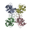
| Title | Crystal structure of Lymphocytic choriomeningitis mammarenavirus endonuclease complexed with catalytic ions | ||||||
 Components Components | RNA-directed RNA polymerase L | ||||||
 Keywords Keywords | TRANSFERASE / Endonuclease / LCMV Arenavirus | ||||||
| Function / homology |  Function and homology information Function and homology informationRNA-templated viral transcription / negative stranded viral RNA replication / cap snatching / virion component / host cell / Hydrolases; Acting on ester bonds / host cell cytoplasm / RNA-directed RNA polymerase / nucleotide binding / hydrolase activity ...RNA-templated viral transcription / negative stranded viral RNA replication / cap snatching / virion component / host cell / Hydrolases; Acting on ester bonds / host cell cytoplasm / RNA-directed RNA polymerase / nucleotide binding / hydrolase activity / RNA-directed RNA polymerase activity / metal ion binding Similarity search - Function | ||||||
| Biological species |  Lymphocytic choriomeningitis mammarenavirus Lymphocytic choriomeningitis mammarenavirus | ||||||
| Method |  X-RAY DIFFRACTION / SYNCHROTRON / MOLECULAR REPLACEMENT / Resolution: 2.43 Å X-RAY DIFFRACTION / SYNCHROTRON / MOLECULAR REPLACEMENT / Resolution: 2.43 Å | ||||||
 Authors Authors | Saez-Ayala, M. / Yekwa, E.L. / Canard, B. / Ferron, F. | ||||||
| Funding support |  France, 1items France, 1items
| ||||||
 Citation Citation | Journal: IUCrJ / Year: 2018 Title: Crystal structures ofLymphocytic choriomeningitis virusendonuclease domain complexed with diketo-acid ligands. Authors: Saez-Ayala, M. / Yekwa, E.L. / Carcelli, M. / Canard, B. / Alvarez, K. / Ferron, F. | ||||||
| History |
|
- Structure visualization
| Structure viewer | Molecule: MolmilJmol/JSmol |
|---|
- Downloads & links
-Download
| PDBx/mmCIF format | 5ltf.cif.gz | 176.7 KB | Display | PDBx/mmCIF format |
|---|---|---|---|---|
| PDB format | pdb5ltf.ent.gz | 139.4 KB | Display | PDB format |
| PDBx/mmJSON format | 5ltf.json.gz | Tree view | PDBx/mmJSON format | |
| Others |  Other downloads Other downloads |
-Validation report
| Arichive directory | https://data.pdbj.org/pub/pdb/validation_reports/lt/5ltfftp://data.pdbj.org/pub/pdb/validation_reports/lt/5ltf | HTTPS FTP |
|---|
-Related structure data
| Related structure data |  5ltnC  5ltsC  5t2tC  3jsbS C: citing same article ( S: Starting model for refinement |
|---|---|
| Similar structure data |


-Links
 PDBj
PDBj- Assembly
| Deposited unit | 
| ||||||||
|---|---|---|---|---|---|---|---|---|---|
| 1 |
| ||||||||
| Unit cell |
|
-Components
| #1: Protein | Mass: 23722.172 Da / Num. of mol.: 2 Source method: isolated from a genetically manipulated source Source: (gene. exp.) Lymphocytic choriomeningitis mammarenavirusGene: L, KUE_IGS320002 / Production host:  References: UniProt: A0A059U381, UniProt: P14240*PLUS, RNA-directed RNA polymerase #2: Chemical |   Mass: 24.305 Da / Num. of mol.: 2 / Source method: obtained synthetically / Formula: Mg Mass: 24.305 Da / Num. of mol.: 2 / Source method: obtained synthetically / Formula: Mg#3: Water | ChemComp-HOH / |  Mass: 18.015 Da / Num. of mol.: 158 / Source method: isolated from a natural source / Formula: H2O Mass: 18.015 Da / Num. of mol.: 158 / Source method: isolated from a natural source / Formula: H2O |
|---|
-Experimental details
-Experiment
| Experiment | Method: X-RAY DIFFRACTION / Number of used crystals: 1 |
|---|
- Sample preparation
| Crystal | Density Matthews: 3.35 Å3/Da / Density % sol: 63.26 % |
|---|---|
| Crystal grow | Temperature: 293.15 K / Method: vapor diffusion, sitting drop Details: sodium citrate 0,1M pH 6,2, Isopropanol 3,5%, MgCl2 1mM |
-Data collection
| Diffraction | Mean temperature: 93 K |
|---|---|
| Diffraction source | Source: SYNCHROTRON / Site: ESRF / Beamline: ID23-1 / Wavelength: 0.9762 Å |
| Detector | Type: DECTRIS PILATUS 6M-F / Detector: PIXEL / Date: Mar 9, 2015 |
| Radiation | Protocol: SINGLE WAVELENGTH / Monochromatic (M) / Laue (L): M / Scattering type: x-ray |
| Radiation wavelength | Wavelength: 0.9762 Å / Relative weight: 1 |
| Reflection | Resolution: 2.43→48.46 Å / Num. obs: 67793 / % possible obs: 96 % / Redundancy: 2.9 % / CC1/2: 0.999 / Rmerge(I) obs: 0.04644 / Net I/σ(I): 13.61 |
| Reflection shell | Resolution: 2.43→2.52 Å / Redundancy: 2.9 % / CC1/2: 0.586 / % possible all: 96 |
- Processing
| Software |
| |||||||||||||||||||||||||||||||||||||||||||||||||||||||||||||||||||||||||||||||||||||||||||||||||||||||||
|---|---|---|---|---|---|---|---|---|---|---|---|---|---|---|---|---|---|---|---|---|---|---|---|---|---|---|---|---|---|---|---|---|---|---|---|---|---|---|---|---|---|---|---|---|---|---|---|---|---|---|---|---|---|---|---|---|---|---|---|---|---|---|---|---|---|---|---|---|---|---|---|---|---|---|---|---|---|---|---|---|---|---|---|---|---|---|---|---|---|---|---|---|---|---|---|---|---|---|---|---|---|---|---|---|---|---|
| Refinement | Method to determine structure: MOLECULAR REPLACEMENT Starting model: 3JSB Resolution: 2.43→48.46 Å / SU ML: 0.32 / Cross valid method: FREE R-VALUE / σ(F): 0 / Phase error: 25.97
| |||||||||||||||||||||||||||||||||||||||||||||||||||||||||||||||||||||||||||||||||||||||||||||||||||||||||
| Solvent computation | Shrinkage radii: 0.9 Å / VDW probe radii: 1.11 Å | |||||||||||||||||||||||||||||||||||||||||||||||||||||||||||||||||||||||||||||||||||||||||||||||||||||||||
| Refinement step | Cycle: LAST / Resolution: 2.43→48.46 Å
| |||||||||||||||||||||||||||||||||||||||||||||||||||||||||||||||||||||||||||||||||||||||||||||||||||||||||
| Refine LS restraints |
| |||||||||||||||||||||||||||||||||||||||||||||||||||||||||||||||||||||||||||||||||||||||||||||||||||||||||
| LS refinement shell |
| |||||||||||||||||||||||||||||||||||||||||||||||||||||||||||||||||||||||||||||||||||||||||||||||||||||||||
| Refinement TLS params. | Method: refined / Origin x: -39.0485 Å / Origin y: -2.5774 Å / Origin z: -3.0292 Å
| |||||||||||||||||||||||||||||||||||||||||||||||||||||||||||||||||||||||||||||||||||||||||||||||||||||||||
| Refinement TLS group | Selection details: all |
