 Movie
Movie Controller


+ Open data

- Basic information
| Entry | Database: PDB / ID: 5lsp | ||||||
|---|---|---|---|---|---|---|---|
| Title | 107_A07 Fab in complex with fragment of the Met receptor | ||||||
 Components Components |
| ||||||
 Keywords Keywords | TRANSFERASE / Fab / beta propeller / Ig-like domain / cell signalling / Met receptor / antibody binding | ||||||
| Function / homology |  Function and homology information Function and homology informationnegative regulation of guanyl-nucleotide exchange factor activity / hepatocyte growth factor receptor activity / Drug-mediated inhibition of MET activation / MET activates STAT3 / negative regulation of hydrogen peroxide-mediated programmed cell death / MET Receptor Activation / MET interacts with TNS proteins / endothelial cell morphogenesis / semaphorin receptor activity / MET receptor recycling ...negative regulation of guanyl-nucleotide exchange factor activity / hepatocyte growth factor receptor activity / Drug-mediated inhibition of MET activation / MET activates STAT3 / negative regulation of hydrogen peroxide-mediated programmed cell death / MET Receptor Activation / MET interacts with TNS proteins / endothelial cell morphogenesis / semaphorin receptor activity / MET receptor recycling / pancreas development / MET activates PTPN11 / MET activates RAP1 and RAC1 / Sema4D mediated inhibition of cell attachment and migration / MET activates PI3K/AKT signaling / positive regulation of endothelial cell chemotaxis / negative regulation of stress fiber assembly / MET activates PTK2 signaling / positive chemotaxis / branching morphogenesis of an epithelial tube / negative regulation of Rho protein signal transduction / negative regulation of thrombin-activated receptor signaling pathway / semaphorin-plexin signaling pathway / establishment of skin barrier / Regulation of MITF-M-dependent genes involved in cell cycle and proliferation / MET activates RAS signaling / MECP2 regulates neuronal receptors and channels / positive regulation of microtubule polymerization / negative regulation of autophagy / cell surface receptor protein tyrosine kinase signaling pathway / basal plasma membrane / molecular function activator activity / InlB-mediated entry of Listeria monocytogenes into host cell / excitatory postsynaptic potential / liver development / receptor protein-tyrosine kinase / Negative regulation of MET activity / neuron differentiation / Constitutive Signaling by Aberrant PI3K in Cancer / PIP3 activates AKT signaling / PI5P, PP2A and IER3 Regulate PI3K/AKT Signaling / RAF/MAP kinase cascade / protein tyrosine kinase activity / protein phosphatase binding / cell surface receptor signaling pathway / receptor complex / postsynapse / cell surface / signal transduction / positive regulation of transcription by RNA polymerase II / extracellular region / ATP binding / identical protein binding / membrane / plasma membrane Similarity search - Function | ||||||
| Biological species |  Homo sapiens (human) Homo sapiens (human) | ||||||
| Method |  X-RAY DIFFRACTION / SYNCHROTRON / MOLECULAR REPLACEMENT / Resolution: 2.605 Å X-RAY DIFFRACTION / SYNCHROTRON / MOLECULAR REPLACEMENT / Resolution: 2.605 Å | ||||||
 Authors Authors | DiCara, D. / Chirgadze, D.Y. / Pope, A. / Karatt-Vellatt, A. / Winter, A. / van den Heuvel, J. / Gherardi, E. / McCafferty, J. | ||||||
 Citation Citation | Journal: Sci Rep / Year: 2017 Title: Characterization and structural determination of a new anti-MET function-blocking antibody with binding epitope distinct from the ligand binding domain. Authors: DiCara, D.M. / Chirgadze, D.Y. / Pope, A.R. / Karatt-Vellatt, A. / Winter, A. / Slavny, P. / van den Heuvel, J. / Parthiban, K. / Holland, J. / Packman, L.C. / Mavria, G. / Hoffmann, J. / ...Authors: DiCara, D.M. / Chirgadze, D.Y. / Pope, A.R. / Karatt-Vellatt, A. / Winter, A. / Slavny, P. / van den Heuvel, J. / Parthiban, K. / Holland, J. / Packman, L.C. / Mavria, G. / Hoffmann, J. / Birchmeier, W. / Gherardi, E. / McCafferty, J. | ||||||
| History |
|
- Structure visualization
| Structure viewer | Molecule: MolmilJmol/JSmol |
|---|
- Downloads & links
-Download
| PDBx/mmCIF format | 5lsp.cif.gz | 263.8 KB | Display | PDBx/mmCIF format |
|---|---|---|---|---|
| PDB format | pdb5lsp.ent.gz | 209.1 KB | Display | PDB format |
| PDBx/mmJSON format | 5lsp.json.gz | Tree view | PDBx/mmJSON format | |
| Others |  Other downloads Other downloads |
-Validation report
| Summary document | 5lsp_validation.pdf.gz | 505.3 KB | Display | wwPDB validaton report |
|---|---|---|---|---|
| Full document | 5lsp_full_validation.pdf.gz | 518.7 KB | Display | |
| Data in XML | 5lsp_validation.xml.gz | 52.7 KB | Display | |
| Data in CIF | 5lsp_validation.cif.gz | 68.4 KB | Display | |
| Arichive directory | https://data.pdbj.org/pub/pdb/validation_reports/ls/5lspftp://data.pdbj.org/pub/pdb/validation_reports/ls/5lsp | HTTPS FTP |
-Related structure data
| Similar structure data |
|---|


-Links
 PDBj
PDBj


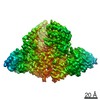
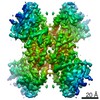
- Assembly
| Deposited unit | 
| ||||||||
|---|---|---|---|---|---|---|---|---|---|
| 1 |
| ||||||||
| Unit cell |
|
-Components
-Hepatocyte growth factor ... , 2 types, 4 molecules APXY
| #1: Protein | Mass: 25592.197 Da / Num. of mol.: 2 Source method: isolated from a genetically manipulated source Source: (gene. exp.) Homo sapiens (human) / Gene: MET / Plasmid: pA71d / Cell line (production host): Lec3.2.8.1 / Production host:   Cricetulus griseus (Chinese hamster) Cricetulus griseus (Chinese hamster)References: UniProt: P08581, receptor protein-tyrosine kinase #4: Protein/peptide | Mass: 1627.857 Da / Num. of mol.: 2 Source method: isolated from a genetically manipulated source Source: (gene. exp.) Homo sapiens (human) / Gene: MET / Plasmid: pA71d / Cell line (production host): Lec3.2.8.1 / Production host: Cricetulus griseus (Chinese hamster)References: UniProt: P08581, receptor protein-tyrosine kinase |
|---|
-Antibody , 2 types, 4 molecules HSLT
| #2: Antibody | Mass: 23891.826 Da / Num. of mol.: 2 Source method: isolated from a genetically manipulated source Source: (gene. exp.) Homo sapiens (human) / Cell line (production host): HEK293 / Production host: Homo sapiens (human)#3: Antibody | Mass: 23504.945 Da / Num. of mol.: 2 Source method: isolated from a genetically manipulated source Source: (gene. exp.) Homo sapiens (human) / Cell line (production host): HEK293 / Production host: Homo sapiens (human) |
|---|
-Sugars / Non-polymers , 2 types, 90 molecules 

| #5: Sugar |  Type: D-saccharide, beta linking / Mass: 221.208 Da / Num. of mol.: 2 Type: D-saccharide, beta linking / Mass: 221.208 Da / Num. of mol.: 2Source method: isolated from a genetically manipulated source Formula: C8H15NO6 #6: Water | ChemComp-HOH / | Mass: 18.015 Da / Num. of mol.: 88 / Source method: isolated from a natural source / Formula: H2O |
|---|
-Details
| Has protein modification | Y |
|---|
-Experimental details
-Experiment
| Experiment | Method: X-RAY DIFFRACTION / Number of used crystals: 1 |
|---|
- Sample preparation
| Crystal | Density Matthews: 2.65 Å3/Da / Density % sol: 53.56 % |
|---|---|
| Crystal grow | Temperature: 293 K / Method: vapor diffusion, hanging drop / pH: 8.5 Details: 10% PEG 20,000, 20% PEG 550-MME, 0.1M Trizma/Bicine pH 8.5, 0.03M magnesium chloride, 0.03M calcium chloride |
-Data collection
| Diffraction | Mean temperature: 100 K | ||||||||||||||||||
|---|---|---|---|---|---|---|---|---|---|---|---|---|---|---|---|---|---|---|---|
| Diffraction source | Source: SYNCHROTRON / Site: ESRF  / Beamline: ID29 / Wavelength: 0.91376 Å / Beamline: ID29 / Wavelength: 0.91376 Å | ||||||||||||||||||
| Detector | Type: PSI PILATUS 6M / Detector: PIXEL / Date: May 29, 2012 | ||||||||||||||||||
| Radiation | Monochromator: Si(311) / Protocol: SINGLE WAVELENGTH / Monochromatic (M) / Laue (L): M / Scattering type: x-ray | ||||||||||||||||||
| Radiation wavelength | Wavelength: 0.91376 Å / Relative weight: 1 | ||||||||||||||||||
| Reflection | Resolution: 2.6→48.95 Å / Num. obs: 49099 / % possible obs: 99 % / Redundancy: 5.5 % / Biso Wilson estimate: 50.2 Å2 / Rmerge(I) obs: 0.11 / Rsym value: 0.11 / Net I/σ(I): 11.7 | ||||||||||||||||||
| Reflection shell |
|
- Processing
| Software |
| |||||||||||||||||||||||||||||||||||||||||||||||||||||||||||||||||||||||||||||||||||||||||||||||||||||||||
|---|---|---|---|---|---|---|---|---|---|---|---|---|---|---|---|---|---|---|---|---|---|---|---|---|---|---|---|---|---|---|---|---|---|---|---|---|---|---|---|---|---|---|---|---|---|---|---|---|---|---|---|---|---|---|---|---|---|---|---|---|---|---|---|---|---|---|---|---|---|---|---|---|---|---|---|---|---|---|---|---|---|---|---|---|---|---|---|---|---|---|---|---|---|---|---|---|---|---|---|---|---|---|---|---|---|---|
| Refinement | Method to determine structure: MOLECULAR REPLACEMENT / Resolution: 2.605→48.942 Å / SU ML: 0.34 / Cross valid method: THROUGHOUT / σ(F): 1.44 / Phase error: 27.53
| |||||||||||||||||||||||||||||||||||||||||||||||||||||||||||||||||||||||||||||||||||||||||||||||||||||||||
| Solvent computation | Shrinkage radii: 0.9 Å / VDW probe radii: 1.11 Å | |||||||||||||||||||||||||||||||||||||||||||||||||||||||||||||||||||||||||||||||||||||||||||||||||||||||||
| Refinement step | Cycle: LAST / Resolution: 2.605→48.942 Å
| |||||||||||||||||||||||||||||||||||||||||||||||||||||||||||||||||||||||||||||||||||||||||||||||||||||||||
| Refine LS restraints |
| |||||||||||||||||||||||||||||||||||||||||||||||||||||||||||||||||||||||||||||||||||||||||||||||||||||||||
| LS refinement shell |
|
