 Movie
Movie Controller

[English] 日本語
 Yorodumi
- PDB-3v00: Studies of a constitutively active G-alpha subunit provide insigh... -
+ Open data

- Basic information
| Entry | Database: PDB / ID: 3v00 | ||||||
|---|---|---|---|---|---|---|---|
| Title | Studies of a constitutively active G-alpha subunit provide insights into the mechanism of G protein activation. | ||||||
 Components Components | Guanine nucleotide-binding protein G(t) subunit alpha-1/ Guanine nucleotide-binding protein G(i) subunit alpha-1 chimeric protein | ||||||
 Keywords Keywords | SIGNALING PROTEIN / GTPase / GTP binding / Transducer / SIGNAL TRANSDUCTION / CELL CYCLE | ||||||
| Function / homology |  Function and homology information Function and homology informationnegative regulation of cyclic-nucleotide phosphodiesterase activity / adenylate cyclase regulator activity / Extra-nuclear estrogen signaling / Adenylate cyclase inhibitory pathway / detection of light stimulus involved in visual perception / G protein-coupled receptor complex / Inactivation, recovery and regulation of the phototransduction cascade / Activation of the phototransduction cascade / negative regulation of synaptic transmission / Adrenaline,noradrenaline inhibits insulin secretion ...negative regulation of cyclic-nucleotide phosphodiesterase activity / adenylate cyclase regulator activity / Extra-nuclear estrogen signaling / Adenylate cyclase inhibitory pathway / detection of light stimulus involved in visual perception / G protein-coupled receptor complex / Inactivation, recovery and regulation of the phototransduction cascade / Activation of the phototransduction cascade / negative regulation of synaptic transmission / Adrenaline,noradrenaline inhibits insulin secretion / ADP signalling through P2Y purinoceptor 12 / GTPase activating protein binding / G alpha (i) signalling events / photoreceptor outer segment membrane / G alpha (i) signalling events / neurotransmitter receptor localization to postsynaptic specialization membrane / acyl binding / phototransduction, visible light / phototransduction / response to light stimulus / adenylate cyclase inhibitor activity / positive regulation of protein localization to cell cortex / photoreceptor inner segment / T cell migration / D2 dopamine receptor binding / adenylate cyclase-inhibiting serotonin receptor signaling pathway / G protein-coupled serotonin receptor binding / cellular response to forskolin / regulation of mitotic spindle organization / chemokine-mediated signaling pathway / response to prostaglandin E / positive regulation of cholesterol biosynthetic process / negative regulation of insulin secretion / G protein-coupled receptor binding / centriolar satellite / G-protein beta/gamma-subunit complex binding / adenylate cyclase-modulating G protein-coupled receptor signaling pathway / adenylate cyclase-inhibiting G protein-coupled receptor signaling pathway / photoreceptor disc membrane / GDP binding / heterotrimeric G-protein complex / G protein activity / midbody / cell cortex / Hydrolases; Acting on acid anhydrides; Acting on GTP to facilitate cellular and subcellular movement / postsynapse / ciliary basal body / G protein-coupled receptor signaling pathway / cell division / GTPase activity / centrosome / protein kinase binding / GTP binding / nucleolus / glutamatergic synapse / magnesium ion binding / Golgi apparatus / protein-containing complex / nucleoplasm / metal ion binding / plasma membrane / cytoplasm / cytosol Similarity search - Function | ||||||
| Biological species |  | ||||||
| Method |  X-RAY DIFFRACTION / SYNCHROTRON / MOLECULAR REPLACEMENT / Resolution: 2.9 Å X-RAY DIFFRACTION / SYNCHROTRON / MOLECULAR REPLACEMENT / Resolution: 2.9 Å | ||||||
 Authors Authors | Singh, G. / Cerione, R.A. | ||||||
 Citation Citation | Journal: Biochemistry / Year: 2012 Title: A constitutively active G-alpha subunit provide insights into the mechanism of G protein activation Authors: Singh, G. / Ramachandran, S. / Cerione, R.A. | ||||||
| History |
|
- Structure visualization
| Structure viewer | Molecule: MolmilJmol/JSmol |
|---|
- Downloads & links
-Download
| PDBx/mmCIF format | 3v00.cif.gz | 228.7 KB | Display | PDBx/mmCIF format |
|---|---|---|---|---|
| PDB format | pdb3v00.ent.gz | 183.8 KB | Display | PDB format |
| PDBx/mmJSON format | 3v00.json.gz | Tree view | PDBx/mmJSON format | |
| Others |  Other downloads Other downloads |
-Validation report
| Arichive directory | https://data.pdbj.org/pub/pdb/validation_reports/v0/3v00ftp://data.pdbj.org/pub/pdb/validation_reports/v0/3v00 | HTTPS FTP |
|---|
-Related structure data
| Related structure data | 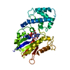 1tagS S: Starting model for refinement |
|---|---|
| Similar structure data |


-Links
 PDBj
PDBj


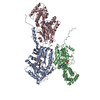
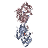
- Assembly


-Components
| #1: Protein | Mass: 40999.656 Da / Num. of mol.: 3 / Fragment: UNP P04695 1-215 & 295-350, UNP P10824 220-298 / Mutation: G56P, K244H and D247N Source method: isolated from a genetically manipulated source Details: T7 LAC PROMOTER Source: (gene. exp.) Gene: GNAT1, GNAT1_BOVIN / Plasmid: PET VECTOR / Production host:  #2: Chemical |   Type: RNA linking / Mass: 443.201 Da / Num. of mol.: 3 / Source method: obtained synthetically / Formula: C10H15N5O11P2 / Comment: GDP, energy-carrying molecule*YM Type: RNA linking / Mass: 443.201 Da / Num. of mol.: 3 / Source method: obtained synthetically / Formula: C10H15N5O11P2 / Comment: GDP, energy-carrying molecule*YM#3: Water | ChemComp-HOH / |  Mass: 18.015 Da / Num. of mol.: 295 / Source method: isolated from a natural source / Formula: H2O Mass: 18.015 Da / Num. of mol.: 295 / Source method: isolated from a natural source / Formula: H2O |
|---|
-Experimental details
-Experiment
| Experiment | Method: X-RAY DIFFRACTION / Number of used crystals: 1 |
|---|
- Sample preparation
| Crystal | Density Matthews: 3.36 Å3/Da / Density % sol: 63.39 % |
|---|---|
| Crystal grow | Temperature: 295 K / Method: vapor diffusion, hanging drop / pH: 6.3 Details: 1.9 M Ammonium Sulphate in 0.05 M Sodium Cacodylate buffer, pH 6.3, VAPOR DIFFUSION, HANGING DROP, temperature 295K |
-Data collection
| Diffraction | Mean temperature: 100 K |
|---|---|
| Diffraction source | Source: SYNCHROTRON / Site: CHESS  / Beamline: A1 / Wavelength: 0.978 Å / Beamline: A1 / Wavelength: 0.978 Å |
| Detector | Type: ADSC QUANTUM 210 / Detector: CCD / Date: Nov 8, 2007 / Details: Mirrors |
| Radiation | Monochromator: Horizontal focusing 5.05 asymmetric cut Si(111) Protocol: SINGLE WAVELENGTH / Monochromatic (M) / Laue (L): M / Scattering type: x-ray |
| Radiation wavelength | Wavelength: 0.978 Å / Relative weight: 1 |
| Reflection | Resolution: 2.7→39.74 Å / Num. all: 47386 / Num. obs: 47006 / % possible obs: 99.2 % / Observed criterion σ(F): 0 / Observed criterion σ(I): 11.3 / Redundancy: 8.6 % / Biso Wilson estimate: 89.2 Å2 / Rmerge(I) obs: 0.08 / Rsym value: 0.08 / Net I/σ(I): 21.253 |
| Reflection shell | Resolution: 2.7→2.8 Å / % possible all: 8.8 |
- Processing
| Software |
| ||||||||||||||||||||||||||||||||||||||||||||||||||||||||||||
|---|---|---|---|---|---|---|---|---|---|---|---|---|---|---|---|---|---|---|---|---|---|---|---|---|---|---|---|---|---|---|---|---|---|---|---|---|---|---|---|---|---|---|---|---|---|---|---|---|---|---|---|---|---|---|---|---|---|---|---|---|---|
| Refinement | Method to determine structure: MOLECULAR REPLACEMENT Starting model: PDB ENTRY 1TAG Resolution: 2.9→39.74 Å / Rfactor Rfree error: 0.005 / Cross valid method: THROUGHOUT / σ(F): 0 / Stereochemistry target values: ENGH & HUBER
| ||||||||||||||||||||||||||||||||||||||||||||||||||||||||||||
| Displacement parameters | Biso mean: 94.5 Å2 | ||||||||||||||||||||||||||||||||||||||||||||||||||||||||||||
| Refine analyze | Luzzati coordinate error free: 0.46 Å / Luzzati sigma a free: 0.56 Å | ||||||||||||||||||||||||||||||||||||||||||||||||||||||||||||
| Refinement step | Cycle: LAST / Resolution: 2.9→39.74 Å
| ||||||||||||||||||||||||||||||||||||||||||||||||||||||||||||
| Refine LS restraints |
|
