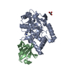
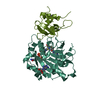
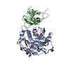
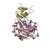
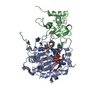
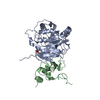
Tumornecrosisfactoralpha-inducedprotein3 / TNF alpha-induced protein 3 / OTU domain-containing protein 7C / Putative DNA-binding protein A20 / ...TNF alpha-induced protein 3 / OTU domain-containing protein 7C / Putative DNA-binding protein A20 / Zinc finger protein A20
Mass: 43514.934 Da / Num. of mol.: 2 Source method: isolated from a genetically manipulated source Source: (gene. exp.) Homo sapiens (human) / Gene: TNFAIP3, OTUD7C / Production host: Escherichia coli (E. coli) References: UniProt: P21580, ubiquitinyl hydrolase 1, Ligases; Forming carbon-nitrogen bonds; Acid-amino-acid ligases (peptide synthases)
#2: Protein
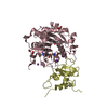
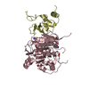
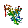
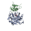
Polyubiquitin-B
Mass: 8558.857 Da / Num. of mol.: 2 / Mutation: G76X Source method: isolated from a genetically manipulated source Details: Gly76 was replaced with propargylamide / Source: (gene. exp.) Homo sapiens (human) / Gene: UBB / Production host: Escherichia coli (E. coli) / References: UniProt: P0CG47
#3: Protein
Tumornecrosisfactoralpha-inducedprotein3 / TNF alpha-induced protein 3 / OTU domain-containing protein 7C / Putative DNA-binding protein A20 / ...TNF alpha-induced protein 3 / OTU domain-containing protein 7C / Putative DNA-binding protein A20 / Zinc finger protein A20
Mass: 43546.930 Da / Num. of mol.: 2 Source method: isolated from a genetically manipulated source Details: Cys103 is oxidized / Source: (gene. exp.) Homo sapiens (human) / Gene: TNFAIP3, OTUD7C / Production host: Escherichia coli (E. coli) References: UniProt: P21580, ubiquitinyl hydrolase 1, Ligases; Forming carbon-nitrogen bonds; Acid-amino-acid ligases (peptide synthases)
Has protein modification
Y
-
Experimental details
-
Experiment
Experiment
Method: X-RAY DIFFRACTION / Number of used crystals: 1
-
Sample preparation
Crystal
Density Matthews: 2.45 Å3/Da / Density % sol: 49.89 %
Crystal grow
Temperature: 287 K / Method: vapor diffusion, sitting drop Details: 0.1 M MES/imidazole (pH 6.5), 7% (w/v) PEG 8K, 20% ethylene glycol
In the structure databanks used in Yorodumi, some data are registered as the other names, "COVID-19 virus" and "2019-nCoV". Here are the details of the virus and the list of structure data.
Jan 31, 2019. EMDB accession codes are about to change! (news from PDBe EMDB page)
EMDB accession codes are about to change! (news from PDBe EMDB page)
The allocation of 4 digits for EMDB accession codes will soon come to an end. Whilst these codes will remain in use, new EMDB accession codes will include an additional digit and will expand incrementally as the available range of codes is exhausted. The current 4-digit format prefixed with “EMD-” (i.e. EMD-XXXX) will advance to a 5-digit format (i.e. EMD-XXXXX), and so on. It is currently estimated that the 4-digit codes will be depleted around Spring 2019, at which point the 5-digit format will come into force.
The EM Navigator/Yorodumi systems omit the EMD- prefix.
Related info.:Q: What is EMD? / ID/Accession-code notation in Yorodumi/EM Navigator
Yorodumi is a browser for structure data from EMDB, PDB, SASBDB, etc.
This page is also the successor to EM Navigator detail page, and also detail information page/front-end page for Omokage search.
The word "yorodu" (or yorozu) is an old Japanese word meaning "ten thousand". "mi" (miru) is to see.
Related info.:EMDB / PDB / SASBDB / Comparison of 3 databanks / Yorodumi Search / Aug 31, 2016. New EM Navigator & Yorodumi / Yorodumi Papers / Jmol/JSmol / Function and homology information / Changes in new EM Navigator and Yorodumi
 Movie
Movie Controller


 Open data
 Basic information
 Components
 Keywords
 Function and homology information
 Homo sapiens (human)
 X-RAY DIFFRACTION /
  Authors
 United Kingdom, 2items
  Citation
 Structure visualization
 Downloads & links
 Other downloads


 PDBj
PDBj


 Assembly


 Sample preparation
 / Beamline: ID29 / Wavelength: 0.96863 Å
 Processing