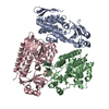
Entry Database : PDB / ID : 5lq4Title The Structure of ThcOx, the First Oxidase Protein from the Cyanobactin Pathways (CyaGox) x 2 Keywords / Function / homology Function Domain/homology Component
/ / / / / / / / / / / / / / / / / / / / / Biological species Cyanothece sp. Cyanothece sp. PCC 7425 (bacteria)Method / / / Resolution : 2.65 Å Authors Bent, A.F. / Wagner, A. / Naismith, J.H. Funding support Organization Grant number Country Biotechnology and Biological Sciences Research Council BB/K015508/1 European Research Council 339367
Journal : Acta Crystallogr D Struct Biol / Year : 2016Title : Structure of the cyanobactin oxidase ThcOx from Cyanothece sp. PCC 7425, the first structure to be solved at Diamond Light Source beamline I23 by means of S-SAD.Authors : Bent, A.F. / Mann, G. / Houssen, W.E. / Mykhaylyk, V. / Duman, R. / Thomas, L. / Jaspars, M. / Wagner, A. / Naismith, J.H. History Deposition Aug 16, 2016 Deposition site / Processing site Revision 1.0 Nov 9, 2016 Provider / Type Revision 1.1 Nov 23, 2016 Group Revision 1.2 Aug 30, 2017 Group / Category / Item Revision 1.3 May 8, 2024 Group / Database references / Refinement descriptionCategory chem_comp_atom / chem_comp_bond ... chem_comp_atom / chem_comp_bond / database_2 / struct_ncs_dom_lim Item _database_2.pdbx_DOI / _database_2.pdbx_database_accession ... _database_2.pdbx_DOI / _database_2.pdbx_database_accession / _struct_ncs_dom_lim.beg_auth_comp_id / _struct_ncs_dom_lim.beg_label_asym_id / _struct_ncs_dom_lim.beg_label_comp_id / _struct_ncs_dom_lim.beg_label_seq_id / _struct_ncs_dom_lim.end_auth_comp_id / _struct_ncs_dom_lim.end_label_asym_id / _struct_ncs_dom_lim.end_label_comp_id / _struct_ncs_dom_lim.end_label_seq_id
Show all Show less
 Movie
Movie Controller

 Yorodumi
 Open data
 Basic information
 Components
 Keywords
 Function and homology information
 Cyanothece sp. PCC 7425 (bacteria)
 X-RAY DIFFRACTION /
  Authors
 United Kingdom, 2items
  Citation
 Structure visualization
 Downloads & links
 Other downloads
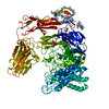


 PDBj
PDBj Assembly
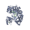
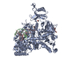
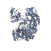
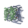

 Mass: 456.344 Da / Num. of mol.: 2
 Mass: 18.015 Da / Num. of mol.: 4 / Source method: isolated from a natural source / Formula: H2O
 Sample preparation
 Processing