[English] 日本語
Yorodumi
- PDB-4mz7: Structural insight into dGTP-dependent activation of tetrameric S... -

+
Open data


ID or keywords:

Loading...

-
Basic information

Entry
Database: PDB / ID: 4mz7
TitleStructural insight into dGTP-dependent activation of tetrameric SAMHD1 deoxynucleoside triphosphate triphosphohydrolase
ComponentsDeoxynucleoside triphosphate triphosphohydrolase SAMHD1
KeywordsHYDROLASE / HD-domain / dNTP Binding / Phosphorylation
Function / homology
Function and homology information


Nucleotide catabolism / Hydrolases; Acting on ester bonds; Triphosphoric-monoester hydrolases / deoxynucleoside triphosphate hydrolase activity / dGTP binding / triphosphoric monoester hydrolase activity / dATP catabolic process / dGTPase activity / tetraspanin-enriched microdomain / dGTP catabolic process / DNA strand resection involved in replication fork processing ...Nucleotide catabolism / Hydrolases; Acting on ester bonds; Triphosphoric-monoester hydrolases / deoxynucleoside triphosphate hydrolase activity / dGTP binding / triphosphoric monoester hydrolase activity / dATP catabolic process / dGTPase activity / tetraspanin-enriched microdomain / dGTP catabolic process / DNA strand resection involved in replication fork processing / deoxyribonucleotide catabolic process / regulation of innate immune response / negative regulation of type I interferon-mediated signaling pathway / somatic hypermutation of immunoglobulin genes / RNA nuclease activity / double-strand break repair via homologous recombination / Interferon alpha/beta signaling / single-stranded DNA binding / site of double-strand break / protein homotetramerization / defense response to virus / nucleic acid binding / immune response / innate immune response / DNA damage response / GTP binding / RNA binding / zinc ion binding / nucleoplasm / identical protein binding / nucleus / plasma membrane
Similarity search - Function
Hypothetical protein af1432 / Hypothetical protein af1432 / HD domain profile. / HD domain / HD domain / SAM domain (Sterile alpha motif) / SAM domain profile. / Metal dependent phosphohydrolases with conserved 'HD' motif. / Sterile alpha motif. / HD/PDEase domain ...Hypothetical protein af1432 / Hypothetical protein af1432 / HD domain profile. / HD domain / HD domain / SAM domain (Sterile alpha motif) / SAM domain profile. / Metal dependent phosphohydrolases with conserved 'HD' motif. / Sterile alpha motif. / HD/PDEase domain / Sterile alpha motif domain / Sterile alpha motif/pointed domain superfamily / Orthogonal Bundle / Mainly Alpha
Similarity search - Domain/homology
2'-DEOXYGUANOSINE-5'-TRIPHOSPHATE / 2'-DEOXYADENOSINE 5'-TRIPHOSPHATE / Deoxynucleoside triphosphate triphosphohydrolase SAMHD1
Similarity search - Component
Biological speciesHomo sapiens (human)
MethodX-RAY DIFFRACTION / SYNCHROTRON / MOLECULAR REPLACEMENT / Resolution: 1.8 Å
AuthorsZhu, C. / Gao, W. / Zhao, K. / Qin, X. / Zhang, Y. / Peng, X. / Zhang, L. / Dong, Y. / Zhang, W. / Li, P. ...Zhu, C. / Gao, W. / Zhao, K. / Qin, X. / Zhang, Y. / Peng, X. / Zhang, L. / Dong, Y. / Zhang, W. / Li, P. / Wei, W. / Gong, Y. / Yu, X.F.
CitationJournal: Nat Commun / Year: 2013
Title: Structural insight into dGTP-dependent activation of tetrameric SAMHD1 deoxynucleoside triphosphate triphosphohydrolase
Authors: Zhu, C. / Gao, W. / Zhao, K. / Qin, X. / Zhang, Y. / Peng, X. / Zhang, L. / Dong, Y. / Zhang, W. / Li, P. / Wei, W. / Gong, Y. / Yu, X.F.
History
DepositionSep 29, 2013Deposition site: RCSB / Processing site: PDBJ
Revision 1.0Nov 20, 2013Provider: repository / Type: Initial release
Revision 1.1Dec 18, 2013Group: Database references
Revision 1.2Mar 29, 2017Group: Refinement description
Revision 1.3Nov 8, 2023Group: Data collection / Database references ...Data collection / Database references / Derived calculations / Refinement description
Category: chem_comp_atom / chem_comp_bond ...chem_comp_atom / chem_comp_bond / database_2 / pdbx_initial_refinement_model / pdbx_struct_conn_angle / struct_conn / struct_ref_seq_dif / struct_site
Item: _database_2.pdbx_DOI / _database_2.pdbx_database_accession ..._database_2.pdbx_DOI / _database_2.pdbx_database_accession / _pdbx_struct_conn_angle.ptnr1_auth_asym_id / _pdbx_struct_conn_angle.ptnr1_auth_comp_id / _pdbx_struct_conn_angle.ptnr1_auth_seq_id / _pdbx_struct_conn_angle.ptnr1_label_asym_id / _pdbx_struct_conn_angle.ptnr1_label_atom_id / _pdbx_struct_conn_angle.ptnr1_label_comp_id / _pdbx_struct_conn_angle.ptnr1_label_seq_id / _pdbx_struct_conn_angle.ptnr2_auth_asym_id / _pdbx_struct_conn_angle.ptnr2_auth_comp_id / _pdbx_struct_conn_angle.ptnr2_auth_seq_id / _pdbx_struct_conn_angle.ptnr2_label_asym_id / _pdbx_struct_conn_angle.ptnr2_label_atom_id / _pdbx_struct_conn_angle.ptnr2_label_comp_id / _pdbx_struct_conn_angle.ptnr3_auth_asym_id / _pdbx_struct_conn_angle.ptnr3_auth_comp_id / _pdbx_struct_conn_angle.ptnr3_auth_seq_id / _pdbx_struct_conn_angle.ptnr3_label_asym_id / _pdbx_struct_conn_angle.ptnr3_label_atom_id / _pdbx_struct_conn_angle.ptnr3_label_comp_id / _pdbx_struct_conn_angle.ptnr3_label_seq_id / _pdbx_struct_conn_angle.value / _struct_conn.pdbx_dist_value / _struct_conn.ptnr1_auth_asym_id / _struct_conn.ptnr1_auth_comp_id / _struct_conn.ptnr1_auth_seq_id / _struct_conn.ptnr1_label_asym_id / _struct_conn.ptnr1_label_atom_id / _struct_conn.ptnr1_label_comp_id / _struct_conn.ptnr1_label_seq_id / _struct_conn.ptnr2_auth_asym_id / _struct_conn.ptnr2_auth_comp_id / _struct_conn.ptnr2_auth_seq_id / _struct_conn.ptnr2_label_asym_id / _struct_conn.ptnr2_label_atom_id / _struct_conn.ptnr2_label_comp_id / _struct_ref_seq_dif.details / _struct_site.pdbx_auth_asym_id / _struct_site.pdbx_auth_comp_id / _struct_site.pdbx_auth_seq_id

-
Structure visualization

Structure viewerMolecule:
MolmilJmol/JSmol

Downloads & links

-
Assembly

Deposited unit
A: Deoxynucleoside triphosphate triphosphohydrolase SAMHD1
B: Deoxynucleoside triphosphate triphosphohydrolase SAMHD1
hetero molecules


Theoretical massNumber of molelcules
Total (without water)128,17912
Polymers124,9722
Non-polymers3,20710
Water14,754819
1
A: Deoxynucleoside triphosphate triphosphohydrolase SAMHD1
B: Deoxynucleoside triphosphate triphosphohydrolase SAMHD1
hetero molecules

A: Deoxynucleoside triphosphate triphosphohydrolase SAMHD1
B: Deoxynucleoside triphosphate triphosphohydrolase SAMHD1
hetero molecules


Theoretical massNumber of molelcules
Total (without water)256,35824
Polymers249,9454
Non-polymers6,41320
Water724
TypeNameSymmetry operationNumber
identity operation1_555x,y,z1
crystal symmetry operation2_858-x+3,y,-z+31
Buried area30150 Å2
ΔGint-202 kcal/mol
Surface area70970 Å2
MethodPISA
Unit cell
Length a, b, c (Å)147.404, 103.608, 92.134
Angle α, β, γ (deg.)90.00, 121.98, 90.00
Int Tables number5
Space group name H-MC121

-
Components

-
Protein , 1 types, 2 molecules AB

#1: Protein Deoxynucleoside triphosphate triphosphohydrolase SAMHD1 / dNTPase / Dendritic cell-derived IFNG-induced protein / DCIP / Monocyte protein 5 / MOP-5 / SAM ...dNTPase / Dendritic cell-derived IFNG-induced protein / DCIP / Monocyte protein 5 / MOP-5 / SAM domain and HD domain-containing protein 1


Mass: 62486.184 Da / Num. of mol.: 2 / Fragment: UNP residues 109-626 / Mutation: C266Y
Source method: isolated from a genetically manipulated source
Source: (gene. exp.) Homo sapiens (human) / Gene: SAMHD1, MOP5 / Production host: Escherichia coli (E. coli)
References: UniProt: Q9Y3Z3, Hydrolases; Acting on ester bonds; Triphosphoric-monoester hydrolases

-
Non-polymers , 5 types, 829 molecules

#2: Chemical ChemComp-ZN / ZINC ION


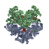
Mass: 65.409 Da / Num. of mol.: 2 / Source method: obtained synthetically / Formula: Zn
#3: Chemical
ChemComp-DGT / 2'-DEOXYGUANOSINE-5'-TRIPHOSPHATE / Deoxyguanosine triphosphate


Mass: 507.181 Da / Num. of mol.: 5 / Source method: obtained synthetically / Formula: C10H16N5O13P3
#4: Chemical ChemComp-MG / MAGNESIUM ION


Mass: 24.305 Da / Num. of mol.: 2 / Source method: obtained synthetically / Formula: Mg
#5: Chemical ChemComp-DTP / 2'-DEOXYADENOSINE 5'-TRIPHOSPHATE / Deoxyadenosine triphosphate


Mass: 491.182 Da / Num. of mol.: 1 / Source method: obtained synthetically / Formula: C10H16N5O12P3
#6: Water ChemComp-HOH / water / Water


Mass: 18.015 Da / Num. of mol.: 819 / Source method: isolated from a natural source / Formula: H2O

-
Experimental details

-
Experiment

ExperimentMethod: X-RAY DIFFRACTION / Number of used crystals: 1

-
Sample preparation

CrystalDensity Matthews: 2.39 Å3/Da / Density % sol: 48.48 %
Crystal growTemperature: 289 K / Method: vapor diffusion, sitting drop / pH: 6.5
Details: 0.1M MES (pH 6.5), 25%(w/v) polyethylene glycol monomethyle ether 550, VAPOR DIFFUSION, SITTING DROP, temperature 289K

-
Data collection

DiffractionMean temperature: 100 K
Diffraction sourceSource: SYNCHROTRON / Site: Photon Factory / Beamline: BL-17A / Wavelength: 0.98 Å
DetectorType: ADSC QUANTUM 270 / Detector: CCD / Date: Jun 19, 2013
RadiationMonochromator: Numerical link type Si(111) double crystal monochromator
Protocol: SINGLE WAVELENGTH / Monochromatic (M) / Laue (L): M / Scattering type: x-ray
Radiation wavelengthWavelength: 0.98 Å / Relative weight: 1
ReflectionResolution: 1.8→50 Å / Num. obs: 108184 / % possible obs: 100 % / Observed criterion σ(F): 1.5 / Observed criterion σ(I): 1.5 / Redundancy: 6.4 % / Biso Wilson estimate: 20.1 Å2 / Rmerge(I) obs: 0.047 / Net I/σ(I): 27.2
Reflection shellResolution: 1.8→1.83 Å / Redundancy: 6.3 % / Rmerge(I) obs: 0.617 / Mean I/σ(I) obs: 2.4 / % possible all: 100

-
Processing

Software
NameVersionClassification
HKL-2000data collection
PHASESphasing
REFMAC5.6.0117refinement
HKL-2000data reduction
HKL-2000data scaling
RefinementMethod to determine structure: MOLECULAR REPLACEMENT
Starting model: PDB ENTRY 3U1N
Resolution: 1.8→50 Å / Cor.coef. Fo:Fc: 0.948 / Cor.coef. Fo:Fc free: 0.93 / Cross valid method: THROUGHOUT / σ(F): 1 / σ(I): 1 / ESU R: 0.135 / ESU R Free: 0.125 / Stereochemistry target values: MAXIMUM LIKELIHOOD
RfactorNum. reflection% reflectionSelection details
Rfree0.22572 5289 5 %RANDOM
Rwork0.19425 ---
all0.196 103173 --
obs0.19582 100284 97.2 %-
Solvent computationIon probe radii: 0.8 Å / Shrinkage radii: 0.8 Å / VDW probe radii: 1.2 Å / Solvent model: MASK
Displacement parametersBiso mean: 22.609 Å2
Baniso -1Baniso -2Baniso -3
1-0.09 Å20 Å2-0.24 Å2
2---0.09 Å20 Å2
3----0.26 Å2
Refinement stepCycle: LAST / Resolution: 1.8→50 Å
ProteinNucleic acidLigandSolventTotal
Num. atoms7974 0 189 819 8982
Refine LS restraints
Refine-IDTypeDev idealDev ideal targetNumber
X-RAY DIFFRACTIONr_bond_refined_d0.0090.028403
X-RAY DIFFRACTIONr_bond_other_d
X-RAY DIFFRACTIONr_angle_refined_deg1.2851.98111399
X-RAY DIFFRACTIONr_angle_other_deg
X-RAY DIFFRACTIONr_dihedral_angle_1_deg5.0555978
X-RAY DIFFRACTIONr_dihedral_angle_2_deg38.73423.995418
X-RAY DIFFRACTIONr_dihedral_angle_3_deg14.685151477
X-RAY DIFFRACTIONr_dihedral_angle_4_deg14.7171554
X-RAY DIFFRACTIONr_chiral_restr0.0870.21192
X-RAY DIFFRACTIONr_gen_planes_refined0.0060.0216350
X-RAY DIFFRACTIONr_gen_planes_other
X-RAY DIFFRACTIONr_nbd_refined
X-RAY DIFFRACTIONr_nbd_other
X-RAY DIFFRACTIONr_nbtor_refined
X-RAY DIFFRACTIONr_nbtor_other
X-RAY DIFFRACTIONr_xyhbond_nbd_refined
X-RAY DIFFRACTIONr_xyhbond_nbd_other
X-RAY DIFFRACTIONr_metal_ion_refined
X-RAY DIFFRACTIONr_metal_ion_other
X-RAY DIFFRACTIONr_symmetry_vdw_refined
X-RAY DIFFRACTIONr_symmetry_vdw_other
X-RAY DIFFRACTIONr_symmetry_hbond_refined
X-RAY DIFFRACTIONr_symmetry_hbond_other
X-RAY DIFFRACTIONr_symmetry_metal_ion_refined
X-RAY DIFFRACTIONr_symmetry_metal_ion_other
X-RAY DIFFRACTIONr_mcbond_it
X-RAY DIFFRACTIONr_mcbond_other
X-RAY DIFFRACTIONr_mcangle_it
X-RAY DIFFRACTIONr_mcangle_other
X-RAY DIFFRACTIONr_scbond_it
X-RAY DIFFRACTIONr_scbond_other
X-RAY DIFFRACTIONr_scangle_it
X-RAY DIFFRACTIONr_scangle_other
X-RAY DIFFRACTIONr_long_range_B_refined
X-RAY DIFFRACTIONr_long_range_B_other
X-RAY DIFFRACTIONr_rigid_bond_restr
X-RAY DIFFRACTIONr_sphericity_free
X-RAY DIFFRACTIONr_sphericity_bonded
LS refinement shellResolution: 1.8→1.847 Å / Rfactor Rfree error: 0 / Total num. of bins used: 20
RfactorNum. reflection% reflection
Rfree0.285 275 -
Rwork0.262 5454 -
all-5729 -
obs--71.75 %

+
About Yorodumi

-
News

-
Feb 9, 2022. New format data for meta-information of EMDB entries

New format data for meta-information of EMDB entries

  • Version 3 of the EMDB header file is now the official format.
  • The previous official version 1.9 will be removed from the archive.

Related info.:EMDB header

External links:wwPDB to switch to version 3 of the EMDB data model

-
Aug 12, 2020. Covid-19 info

Covid-19 info

URL: https://pdbj.org/emnavi/covid19.php

New page: Covid-19 featured information page in EM Navigator.

Related info.:Covid-19 info / Mar 5, 2020. Novel coronavirus structure data

+
Mar 5, 2020. Novel coronavirus structure data

Novel coronavirus structure data

Related info.:Yorodumi Speices / Aug 12, 2020. Covid-19 info

External links:COVID-19 featured content - PDBj / Molecule of the Month (242):Coronavirus Proteases

+
Jan 31, 2019. EMDB accession codes are about to change! (news from PDBe EMDB page)

EMDB accession codes are about to change! (news from PDBe EMDB page)

  • The allocation of 4 digits for EMDB accession codes will soon come to an end. Whilst these codes will remain in use, new EMDB accession codes will include an additional digit and will expand incrementally as the available range of codes is exhausted. The current 4-digit format prefixed with “EMD-” (i.e. EMD-XXXX) will advance to a 5-digit format (i.e. EMD-XXXXX), and so on. It is currently estimated that the 4-digit codes will be depleted around Spring 2019, at which point the 5-digit format will come into force.
  • The EM Navigator/Yorodumi systems omit the EMD- prefix.

Related info.:Q: What is EMD? / ID/Accession-code notation in Yorodumi/EM Navigator

External links:EMDB Accession Codes are Changing Soon! / Contact to PDBj

+
Jul 12, 2017. Major update of PDB

Major update of PDB

  • wwPDB released updated PDB data conforming to the new PDBx/mmCIF dictionary.
  • This is a major update changing the version number from 4 to 5, and with Remediation, in which all the entries are updated.
  • In this update, many items about electron microscopy experimental information are reorganized (e.g. em_software).
  • Now, EM Navigator and Yorodumi are based on the updated data.

External links:wwPDB Remediation / Enriched Model Files Conforming to OneDep Data Standards Now Available in the PDB FTP Archive

-
Yorodumi

Thousand views of thousand structures

  • Yorodumi is a browser for structure data from EMDB, PDB, SASBDB, etc.
  • This page is also the successor to EM Navigator detail page, and also detail information page/front-end page for Omokage search.
  • The word "yorodu" (or yorozu) is an old Japanese word meaning "ten thousand". "mi" (miru) is to see.

Related info.:EMDB / PDB / SASBDB / Comparison of 3 databanks / Yorodumi Search / Aug 31, 2016. New EM Navigator & Yorodumi / Yorodumi Papers / Jmol/JSmol / Function and homology information / Changes in new EM Navigator and Yorodumi

Read more