 Movie
Movie Controller

[English] 日本語
 Yorodumi
- PDB-5ll0: Structure of Polyphosphate Kinase 2 from Francisella tularensis S... -
+ Open data

- Basic information
| Entry | Database: PDB / ID: 5ll0 | ||||||
|---|---|---|---|---|---|---|---|
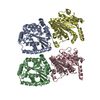
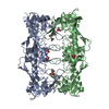
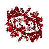
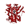
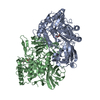
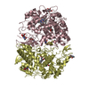
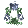
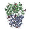
| Title | Structure of Polyphosphate Kinase 2 from Francisella tularensis SCHU S4 with polyphosphate | ||||||
 Components Components | Polyphosphate kinase 2 | ||||||
 Keywords Keywords | TRANSFERASE / Polyphosphate metabolism and nucleotide metabolism / Polyphosphate Kinase 2 enzyme | ||||||
| Function / homology |  Function and homology information Function and homology informationphosphorus metabolic process / Transferases; Transferring phosphorus-containing groups; Phosphotransferases with a phosphate group as acceptor / polyphosphate kinase activity / metal ion binding Similarity search - Function | ||||||
| Biological species |  Francisella tularensis subsp. tularensis (bacteria) Francisella tularensis subsp. tularensis (bacteria) | ||||||
| Method |  X-RAY DIFFRACTION / SYNCHROTRON / MOLECULAR REPLACEMENT / Resolution: 1.96 Å X-RAY DIFFRACTION / SYNCHROTRON / MOLECULAR REPLACEMENT / Resolution: 1.96 Å | ||||||
 Authors Authors | Roach, P.L. / Parnell, A.E. | ||||||
| Funding support |  United States, 1items United States, 1items
| ||||||
 Citation Citation | Journal: Proc. Natl. Acad. Sci. U.S.A. / Year: 2018 Title: Substrate recognition and mechanism revealed by ligand-bound polyphosphate kinase 2 structures. Authors: Parnell, A.E. / Mordhorst, S. / Kemper, F. / Giurrandino, M. / Prince, J.P. / Schwarzer, N.J. / Hofer, A. / Wohlwend, D. / Jessen, H.J. / Gerhardt, S. / Einsle, O. / Oyston, P.C.F. / Andexer, J.N. / Roach, P.L. #1: Journal: Biosci. Rep. / Year: 2016Title: Biochemical and structural characterization of polyphosphate kinase 2 from the intracellular pathogen Francisella tularensis. Authors: Batten, L.E. / Parnell, A.E. / Wells, N.J. / Murch, A.L. / Oyston, P.C. / Roach, P.L. | ||||||
| History |
|
- Structure visualization
| Structure viewer | Molecule: MolmilJmol/JSmol |
|---|
- Downloads & links
-Download
| PDBx/mmCIF format | 5ll0.cif.gz | 395.6 KB | Display | PDBx/mmCIF format |
|---|---|---|---|---|
| PDB format | pdb5ll0.ent.gz | 328.2 KB | Display | PDB format |
| PDBx/mmJSON format | 5ll0.json.gz | Tree view | PDBx/mmJSON format | |
| Others |  Other downloads Other downloads |
-Validation report
| Summary document | 5ll0_validation.pdf.gz | 1.4 MB | Display | wwPDB validaton report |
|---|---|---|---|---|
| Full document | 5ll0_full_validation.pdf.gz | 1.4 MB | Display | |
| Data in XML | 5ll0_validation.xml.gz | 42.6 KB | Display | |
| Data in CIF | 5ll0_validation.cif.gz | 59.4 KB | Display | |
| Arichive directory | https://data.pdbj.org/pub/pdb/validation_reports/ll/5ll0ftp://data.pdbj.org/pub/pdb/validation_reports/ll/5ll0 | HTTPS FTP |
-Related structure data
| Related structure data | 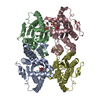 5lc9C 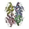 5lcdC 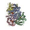 5ld1C 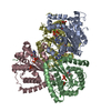 5ldbC 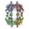 5llbC 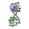 5llfC 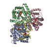 5maqC 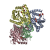 5o6kC 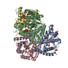 5o6mC 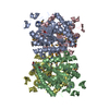 3czqS S: Starting model for refinement C: citing same article ( |
|---|---|
| Similar structure data |

-Links
 PDBj
PDBj- Assembly
| Deposited unit | 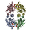
| ||||||||
|---|---|---|---|---|---|---|---|---|---|
| 1 |
| ||||||||
| Unit cell |
|
-Components
| #1: Protein | Mass: 32135.043 Da / Num. of mol.: 4 Source method: isolated from a genetically manipulated source Details: Polyphosphate: Nine phosphates Source: (gene. exp.) Francisella tularensis subsp. tularensis (strain SCHU S4 / Schu 4) (bacteria)Gene: ppk2, FTT_1564, BZ14_1190 / Production host: References: UniProt: Q5NEQ5, ATP-polyphosphate phosphotransferase #2: Chemical | ChemComp-9PI /   Mass: 737.834 Da / Num. of mol.: 4 / Source method: obtained synthetically / Formula: H11O28P9 Mass: 737.834 Da / Num. of mol.: 4 / Source method: obtained synthetically / Formula: H11O28P9#3: Water | ChemComp-HOH / |  Mass: 18.015 Da / Num. of mol.: 612 / Source method: isolated from a natural source / Formula: H2O Mass: 18.015 Da / Num. of mol.: 612 / Source method: isolated from a natural source / Formula: H2O |
|---|
-Experimental details
-Experiment
| Experiment | Method: X-RAY DIFFRACTION / Number of used crystals: 1 |
|---|
- Sample preparation
| Crystal | Density Matthews: 2.44 Å3/Da / Density % sol: 49.66 % |
|---|---|
| Crystal grow | Temperature: 293.15 K / Method: vapor diffusion, hanging drop Details: 5 % Glycerol/PEG 4000, 0.1 M MES/imidazole pH 6.5, 0.15 M Morpheus alcohols, 1 mM polyP, 5 mM MgCl2 |
-Data collection
| Diffraction | Mean temperature: 80 K |
|---|---|
| Diffraction source | Source: SYNCHROTRON / Site: Diamond  / Beamline: I04-1 / Wavelength: 0.92001 Å / Beamline: I04-1 / Wavelength: 0.92001 Å |
| Detector | Type: DECTRIS PILATUS 6M / Detector: PIXEL / Date: Oct 13, 2013 |
| Radiation | Protocol: SINGLE WAVELENGTH / Monochromatic (M) / Laue (L): M / Scattering type: x-ray |
| Radiation wavelength | Wavelength: 0.92001 Å / Relative weight: 1 |
| Reflection | Resolution: 1.96→72.81 Å / Num. obs: 76026 / % possible obs: 97.1 % / Redundancy: 3.3 % / Rmerge(I) obs: 0.06 / Net I/σ(I): 11.7 |
- Processing
| Software |
| ||||||||||||||||||||
|---|---|---|---|---|---|---|---|---|---|---|---|---|---|---|---|---|---|---|---|---|---|
| Refinement | Method to determine structure: MOLECULAR REPLACEMENT Starting model: 3czq Resolution: 1.96→59.142 Å / Cross valid method: FREE R-VALUE
| ||||||||||||||||||||
| Solvent computation | VDW probe radii: 1.11 Å | ||||||||||||||||||||
| Refinement step | Cycle: LAST / Resolution: 1.96→59.142 Å
|
