 Movie
Movie Controller

[English] 日本語
 Yorodumi
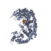
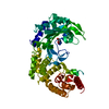
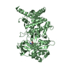
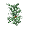
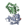
- PDB-1yft: The crystal structure of the catalytic fragment of alanyl-tRNA sy... -
+ Open data

- Basic information
| Entry | Database: PDB / ID: 1yft | ||||||
|---|---|---|---|---|---|---|---|
| Title | The crystal structure of the catalytic fragment of alanyl-tRNA synthetase in complex wtih glycine | ||||||
 Components Components | Alanyl-tRNA synthetase | ||||||
 Keywords Keywords | LIGASE / alpha-beta fold / amino acid binding / helix-loop helix motif | ||||||
| Function / homology |  Function and homology information Function and homology informationalanyl-tRNA aminoacylation / alanine-tRNA ligase / alanine-tRNA ligase activity / aminoacyl-tRNA deacylase activity / tRNA binding / zinc ion binding / ATP binding / cytosol Similarity search - Function | ||||||
| Biological species |   Aquifex aeolicus (bacteria) Aquifex aeolicus (bacteria) | ||||||
| Method |  X-RAY DIFFRACTION / SYNCHROTRON / FOURIER SYNTHESIS / Resolution: 2.23 Å X-RAY DIFFRACTION / SYNCHROTRON / FOURIER SYNTHESIS / Resolution: 2.23 Å | ||||||
 Authors Authors | Swairjo, M.A. / Schimmel, P.R. | ||||||
 Citation Citation | Journal: Proc.Natl.Acad.Sci.USA / Year: 2005 Title: Breaking sieve for steric exclusion of a noncognate amino acid from active site of a tRNA synthetase. Authors: Swairjo, M.A. / Schimmel, P.R. #1: Journal: Mol.Cell / Year: 2004Title: Alanyl-tRNA synthetase crystal structure and design for acceptor-stem recognition. Authors: Swairjo, M.A. / Otero, F.J. / Yang, X.-L. / Lovato, M.A. / Skene, R.J. / McRee, D.E. / Ribas de Pouplana, L. / Schimmel, P. | ||||||
| History |
|
- Structure visualization
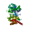
| Structure viewer | Molecule: MolmilJmol/JSmol |
|---|
- Downloads & links
-Download
| PDBx/mmCIF format | 1yft.cif.gz | 106.4 KB | Display | PDBx/mmCIF format |
|---|---|---|---|---|
| PDB format | pdb1yft.ent.gz | 80.8 KB | Display | PDB format |
| PDBx/mmJSON format | 1yft.json.gz | Tree view | PDBx/mmJSON format | |
| Others |  Other downloads Other downloads |
-Validation report
| Arichive directory | https://data.pdbj.org/pub/pdb/validation_reports/yf/1yftftp://data.pdbj.org/pub/pdb/validation_reports/yf/1yft | HTTPS FTP |
|---|
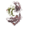
-Related structure data
| Related structure data |  1yfrC 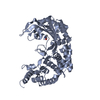 1yfsC  1ygbC  1riqS S: Starting model for refinement C: citing same article ( |
|---|---|
| Similar structure data |


-Links
 PDBj
PDBj


- Assembly
| Deposited unit | 
| ||||||||
|---|---|---|---|---|---|---|---|---|---|
| 1 |
| ||||||||
| 2 | 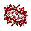
| ||||||||
| Unit cell |
| ||||||||
| Details | the biological unit is the entire asymmetric unit, i.e. one alanyl-tRNa synthetase molecule in complex with one glycine molecule. |
-Components
| #1: Protein | Mass: 53772.246 Da / Num. of mol.: 1 Source method: isolated from a genetically manipulated source Source: (gene. exp.) Aquifex aeolicus (bacteria) / Gene: alaS / Plasmid: PET20B+ / Production host: |
|---|---|
| #2: Chemical | ChemComp-GLY /   Type: peptide linking / Mass: 75.067 Da / Num. of mol.: 1 / Source method: obtained synthetically / Formula: C2H5NO2 Type: peptide linking / Mass: 75.067 Da / Num. of mol.: 1 / Source method: obtained synthetically / Formula: C2H5NO2 |
| #3: Water | ChemComp-HOH /  Mass: 18.015 Da / Num. of mol.: 193 / Source method: isolated from a natural source / Formula: H2O Mass: 18.015 Da / Num. of mol.: 193 / Source method: isolated from a natural source / Formula: H2O |
-Experimental details
-Experiment
| Experiment | Method: X-RAY DIFFRACTION / Number of used crystals: 1 |
|---|
- Sample preparation
| Crystal | Density Matthews: 2.26 Å3/Da / Density % sol: 46 % |
|---|---|
| Crystal grow | Temperature: 298 K / Method: vapor diffusion, sitting drop / pH: 6.2 Details: PEG 5000 mme, NaCL, Tris-Cl, Tris-base, glycine, pH 6.2, VAPOR DIFFUSION, SITTING DROP, temperature 298K |
-Data collection
| Diffraction | Mean temperature: 200 K |
|---|---|
| Diffraction source | Source: SYNCHROTRON / Site: SSRL  / Beamline: BL11-1 / Wavelength: 0.82653 Å / Beamline: BL11-1 / Wavelength: 0.82653 Å |
| Detector | Type: ADSC QUANTUM 315 / Detector: CCD / Date: Jul 16, 2004 |
| Radiation | Monochromator: curved crystal / Protocol: SINGLE WAVELENGTH / Monochromatic (M) / Laue (L): M / Scattering type: x-ray |
| Radiation wavelength | Wavelength: 0.82653 Å / Relative weight: 1 |
| Reflection | Resolution: 2.23→50 Å / Num. obs: 24100 / % possible obs: 99.7 % / Redundancy: 5.4 % / Rmerge(I) obs: 0.083 / Net I/σ(I): 12 |
| Reflection shell | Resolution: 2.23→2.33 Å / Redundancy: 5.4 % / Rmerge(I) obs: 0.44 / % possible all: 100 |
- Processing
| Software |
| |||||||||||||||||||||||||
|---|---|---|---|---|---|---|---|---|---|---|---|---|---|---|---|---|---|---|---|---|---|---|---|---|---|---|
| Refinement | Method to determine structure: FOURIER SYNTHESIS Starting model: 1RIQ Resolution: 2.23→50 Å / Isotropic thermal model: isotropic / Cross valid method: THROUGHOUT / σ(F): 0 / Stereochemistry target values: Engh & Huber
| |||||||||||||||||||||||||
| Displacement parameters | Biso mean: 21.9 Å2 | |||||||||||||||||||||||||
| Refinement step | Cycle: LAST / Resolution: 2.23→50 Å
| |||||||||||||||||||||||||
| Refine LS restraints |
| |||||||||||||||||||||||||
| LS refinement shell | Resolution: 2.23→2.33 Å / Rfactor Rfree error: 0.233 / Total num. of bins used: 8
|
