 Movie
Movie Controller

[English] 日本語
 Yorodumi
- PDB-5o6m: Structure of Polyphosphate Kinase from Meiothermus ruber N121D bo... -
+ Open data

- Basic information
| Entry | Database: PDB / ID: 5o6m | ||||||
|---|---|---|---|---|---|---|---|
| Title | Structure of Polyphosphate Kinase from Meiothermus ruber N121D bound to ATP | ||||||
 Components Components | Polyphosphate:AMP phosphotransferase | ||||||
 Keywords Keywords | TRANSFERASE / Polyphosphate Kinase type 2 class III | ||||||
| Function / homology |  Function and homology information Function and homology informationTransferases; Transferring phosphorus-containing groups; Phosphotransferases with a phosphate group as acceptor / polyphosphate kinase activity / phosphotransferase activity, phosphate group as acceptor / polyphosphate metabolic process / kinase activity Similarity search - Function | ||||||
| Biological species |  Meiothermus ruber H328 (bacteria) Meiothermus ruber H328 (bacteria) | ||||||
| Method |  X-RAY DIFFRACTION / SYNCHROTRON / MOLECULAR REPLACEMENT / Resolution: 2.3 Å X-RAY DIFFRACTION / SYNCHROTRON / MOLECULAR REPLACEMENT / Resolution: 2.3 Å | ||||||
 Authors Authors | Kemper, F. / Gerhardt, S. / Einsle, O. | ||||||
 Citation Citation | Journal: Proc. Natl. Acad. Sci. U.S.A. / Year: 2018 Title: Substrate recognition and mechanism revealed by ligand-bound polyphosphate kinase 2 structures. Authors: Parnell, A.E. / Mordhorst, S. / Kemper, F. / Giurrandino, M. / Prince, J.P. / Schwarzer, N.J. / Hofer, A. / Wohlwend, D. / Jessen, H.J. / Gerhardt, S. / Einsle, O. / Oyston, P.C.F. / Andexer, J.N. / Roach, P.L. | ||||||
| History |
|
- Structure visualization
| Structure viewer | Molecule: MolmilJmol/JSmol |
|---|
- Downloads & links
-Download
| PDBx/mmCIF format | 5o6m.cif.gz | 242.7 KB | Display | PDBx/mmCIF format |
|---|---|---|---|---|
| PDB format | pdb5o6m.ent.gz | 195.4 KB | Display | PDB format |
| PDBx/mmJSON format | 5o6m.json.gz | Tree view | PDBx/mmJSON format | |
| Others |  Other downloads Other downloads |
-Validation report
| Arichive directory | https://data.pdbj.org/pub/pdb/validation_reports/o6/5o6mftp://data.pdbj.org/pub/pdb/validation_reports/o6/5o6m | HTTPS FTP |
|---|
-Related structure data
| Related structure data | 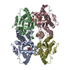 5lc9SC 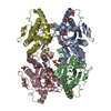 5lcdC 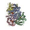 5ld1C 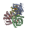 5ldbC 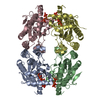 5ll0C 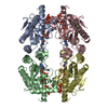 5llbC 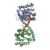 5llfC 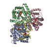 5maqC 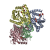 5o6kC S: Starting model for refinement C: citing same article ( |
|---|---|
| Similar structure data |


-Links
 PDBj
PDBj- Assembly
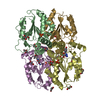
| Deposited unit | 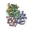
| ||||||||
|---|---|---|---|---|---|---|---|---|---|
| 1 |
| ||||||||
| Unit cell |
|
-Components
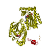
| #1: Protein | Mass: 31607.961 Da / Num. of mol.: 4 / Mutation: N121D Source method: isolated from a genetically manipulated source Source: (gene. exp.) Meiothermus ruber H328 (bacteria) / Gene: MrH_2468 / Production host: #2: Chemical | ChemComp-ATP /   Mass: 507.181 Da / Num. of mol.: 4 / Source method: obtained synthetically / Formula: C10H16N5O13P3 / Comment: ATP, energy-carrying molecule*YM Mass: 507.181 Da / Num. of mol.: 4 / Source method: obtained synthetically / Formula: C10H16N5O13P3 / Comment: ATP, energy-carrying molecule*YM#3: Chemical | ChemComp-PO4 /   Mass: 94.971 Da / Num. of mol.: 22 / Source method: obtained synthetically / Formula: PO4 Mass: 94.971 Da / Num. of mol.: 22 / Source method: obtained synthetically / Formula: PO4#4: Water | ChemComp-HOH / |  Mass: 18.015 Da / Num. of mol.: 294 / Source method: isolated from a natural source / Formula: H2O Mass: 18.015 Da / Num. of mol.: 294 / Source method: isolated from a natural source / Formula: H2O |
|---|
-Experimental details
-Experiment
| Experiment | Method: X-RAY DIFFRACTION / Number of used crystals: 1 |
|---|
- Sample preparation
| Crystal | Density Matthews: 2.55 Å3/Da / Density % sol: 51.74 % |
|---|---|
| Crystal grow | Temperature: 293 K / Method: vapor diffusion, sitting drop / pH: 8.5 Details: 100 mM Tris/HCl pH 8.5 200 mM Li2SO4 27 % (w/v) PEG3350 |
-Data collection
| Diffraction | Mean temperature: 100 K |
|---|---|
| Diffraction source | Source: SYNCHROTRON / Site: SLS  / Beamline: X06DA / Wavelength: 1 Å / Beamline: X06DA / Wavelength: 1 Å |
| Detector | Type: DECTRIS PILATUS 2M / Detector: PIXEL / Date: Apr 9, 2017 / Details: MIRROR |
| Radiation | Monochromator: Si(111) / Protocol: SINGLE WAVELENGTH / Monochromatic (M) / Laue (L): M / Scattering type: x-ray |
| Radiation wavelength | Wavelength: 1 Å / Relative weight: 1 |
| Reflection | Resolution: 2.3→47.56 Å / Num. obs: 58486 / % possible obs: 100 % / Redundancy: 26.4 % / CC1/2: 1 / Rmerge(I) obs: 0.133 / Rpim(I) all: 0.026 / Net I/σ(I): 22.1 |
| Reflection shell | Resolution: 2.3→2.36 Å / Redundancy: 27.6 % / Rmerge(I) obs: 1.659 / Mean I/σ(I) obs: 2.9 / Num. unique obs: 4452 / CC1/2: 0.928 / Rpim(I) all: 0.32 / % possible all: 100 |
- Processing
| Software |
| |||||||||||||||||||||||||||||||||||||||||||||||||||||||||||||||||||||||||||||||||||||||||||||||||||||||||||||||||||||||||||||||||||||||||||||||||||
|---|---|---|---|---|---|---|---|---|---|---|---|---|---|---|---|---|---|---|---|---|---|---|---|---|---|---|---|---|---|---|---|---|---|---|---|---|---|---|---|---|---|---|---|---|---|---|---|---|---|---|---|---|---|---|---|---|---|---|---|---|---|---|---|---|---|---|---|---|---|---|---|---|---|---|---|---|---|---|---|---|---|---|---|---|---|---|---|---|---|---|---|---|---|---|---|---|---|---|---|---|---|---|---|---|---|---|---|---|---|---|---|---|---|---|---|---|---|---|---|---|---|---|---|---|---|---|---|---|---|---|---|---|---|---|---|---|---|---|---|---|---|---|---|---|---|---|---|---|
| Refinement | Method to determine structure: MOLECULAR REPLACEMENT Starting model: 5LC9 Resolution: 2.3→47.532 Å / SU ML: 0.26 / Cross valid method: THROUGHOUT / σ(F): 1.34 / Phase error: 25.59 / Stereochemistry target values: ML
| |||||||||||||||||||||||||||||||||||||||||||||||||||||||||||||||||||||||||||||||||||||||||||||||||||||||||||||||||||||||||||||||||||||||||||||||||||
| Solvent computation | Shrinkage radii: 0.9 Å / VDW probe radii: 1.11 Å / Solvent model: FLAT BULK SOLVENT MODEL | |||||||||||||||||||||||||||||||||||||||||||||||||||||||||||||||||||||||||||||||||||||||||||||||||||||||||||||||||||||||||||||||||||||||||||||||||||
| Refinement step | Cycle: LAST / Resolution: 2.3→47.532 Å
| |||||||||||||||||||||||||||||||||||||||||||||||||||||||||||||||||||||||||||||||||||||||||||||||||||||||||||||||||||||||||||||||||||||||||||||||||||
| Refine LS restraints |
| |||||||||||||||||||||||||||||||||||||||||||||||||||||||||||||||||||||||||||||||||||||||||||||||||||||||||||||||||||||||||||||||||||||||||||||||||||
| LS refinement shell |
|
