 Movie
Movie Controller

[English] 日本語
 Yorodumi
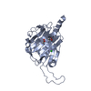
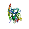
- PDB-5lk5: Crystal structure of the globular domain of human calreticulin mu... -
+ Open data

- Basic information
| Entry | Database: PDB / ID: 5lk5 | ||||||
|---|---|---|---|---|---|---|---|
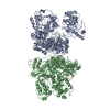
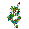
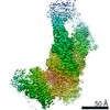
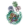
| Title | Crystal structure of the globular domain of human calreticulin mutant D71K | ||||||
 Components Components | Calreticulin,Calreticulin | ||||||
 Keywords Keywords | calcium-binding protein | ||||||
| Function / homology |  Function and homology information Function and homology informationresponse to biphenyl / Calnexin/calreticulin cycle / cytolytic granule / nuclear receptor-mediated glucocorticoid signaling pathway / Assembly of Viral Components at the Budding Site / positive regulation of dendritic cell chemotaxis / ATF6 (ATF6-alpha) activates chaperone genes / negative regulation of trophoblast cell migration / cortical granule / regulation of meiotic nuclear division ...response to biphenyl / Calnexin/calreticulin cycle / cytolytic granule / nuclear receptor-mediated glucocorticoid signaling pathway / Assembly of Viral Components at the Budding Site / positive regulation of dendritic cell chemotaxis / ATF6 (ATF6-alpha) activates chaperone genes / negative regulation of trophoblast cell migration / cortical granule / regulation of meiotic nuclear division / cellular response to electrical stimulus / : / negative regulation of retinoic acid receptor signaling pathway / complement component C1q complex binding / endoplasmic reticulum quality control compartment / protein folding in endoplasmic reticulum / sarcoplasmic reticulum lumen / response to peptide / negative regulation of intracellular steroid hormone receptor signaling pathway / nuclear export signal receptor activity / cardiac muscle cell differentiation / cortical actin cytoskeleton organization / response to glycoside / Scavenging by Class A Receptors / Scavenging by Class F Receptors / nuclear androgen receptor binding / cellular response to lithium ion / negative regulation of neuron differentiation / response to testosterone / smooth endoplasmic reticulum / hormone binding / molecular sequestering activity / protein localization to nucleus / peptide binding / ERAD pathway / endocytic vesicle lumen / positive regulation of cell cycle / positive regulation of substrate adhesion-dependent cell spreading / endoplasmic reticulum-Golgi intermediate compartment membrane / protein export from nucleus / protein folding chaperone / positive regulation of endothelial cell migration / positive regulation of phagocytosis / acrosomal vesicle / Antigen Presentation: Folding, assembly and peptide loading of class I MHC / lumenal side of endoplasmic reticulum membrane / peptide antigen assembly with MHC class I protein complex / MHC class I peptide loading complex / protein maturation / cellular response to virus / positive regulation of non-canonical NF-kappaB signal transduction / integrin binding / phagocytic vesicle membrane / intracellular calcium ion homeostasis / cellular senescence / : / response to estradiol / nuclear envelope / carbohydrate binding / protein folding / protein-folding chaperone binding / extracellular matrix / ER-Phagosome pathway / spermatogenesis / regulation of apoptotic process / negative regulation of translation / protein stabilization / postsynapse / ribosome / iron ion binding / response to xenobiotic stimulus / endoplasmic reticulum lumen / external side of plasma membrane / focal adhesion / negative regulation of DNA-templated transcription / mRNA binding / calcium ion binding / positive regulation of cell population proliferation / ubiquitin protein ligase binding / positive regulation of gene expression / regulation of DNA-templated transcription / endoplasmic reticulum membrane / perinuclear region of cytoplasm / glutamatergic synapse / cell surface / negative regulation of transcription by RNA polymerase II / endoplasmic reticulum / mitochondrion / : / DNA binding / RNA binding / extracellular exosome / extracellular region / zinc ion binding / membrane / nucleus / cytoplasm / cytosol Similarity search - Function | ||||||
| Biological species |  Homo sapiens (human) Homo sapiens (human) | ||||||
| Method |  X-RAY DIFFRACTION / SYNCHROTRON / Resolution: 2.3 Å X-RAY DIFFRACTION / SYNCHROTRON / Resolution: 2.3 Å | ||||||
 Authors Authors | Gaboriaud, C. / Cioci, G. | ||||||
 Citation Citation | Journal: IUCrJ / Year: 2016 Title: Structures of parasite calreticulins provide insights into their flexibility and dual carbohydrate/peptide-binding properties. Authors: Moreau, C. / Cioci, G. / Iannello, M. / Laffly, E. / Chouquet, A. / Ferreira, A. / Thielens, N.M. / Gaboriaud, C. #1: Journal: PLoS ONE / Year: 2011Title: X-ray structure of the human calreticulin globular domain reveals a peptide-binding area and suggests a multi-molecular mechanism. Authors: Chouquet, A. / Paidassi, H. / Ling, W.L. / Frachet, P. / Houen, G. / Arlaud, G.J. / Gaboriaud, C. | ||||||
| History |
|
- Structure visualization
| Structure viewer | Molecule: MolmilJmol/JSmol |
|---|
- Downloads & links
-Download
| PDBx/mmCIF format | 5lk5.cif.gz | 529.1 KB | Display | PDBx/mmCIF format |
|---|---|---|---|---|
| PDB format | pdb5lk5.ent.gz | 430.5 KB | Display | PDB format |
| PDBx/mmJSON format | 5lk5.json.gz | Tree view | PDBx/mmJSON format | |
| Others |  Other downloads Other downloads |
-Validation report
| Arichive directory | https://data.pdbj.org/pub/pdb/validation_reports/lk/5lk5ftp://data.pdbj.org/pub/pdb/validation_reports/lk/5lk5 | HTTPS FTP |
|---|
-Related structure data


-Links
 PDBj
PDBj


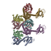
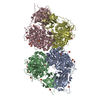
- Assembly
| Deposited unit | 
| |||||||||||||||||||||||||||||||||||||||||||||||||||||||||||||||||||||||||||||||||||||||||||||||||||||||||||||||||||||||||||||||||||||||||||||||||||||||||||||||||||||||||||||||||||||||||||||
|---|---|---|---|---|---|---|---|---|---|---|---|---|---|---|---|---|---|---|---|---|---|---|---|---|---|---|---|---|---|---|---|---|---|---|---|---|---|---|---|---|---|---|---|---|---|---|---|---|---|---|---|---|---|---|---|---|---|---|---|---|---|---|---|---|---|---|---|---|---|---|---|---|---|---|---|---|---|---|---|---|---|---|---|---|---|---|---|---|---|---|---|---|---|---|---|---|---|---|---|---|---|---|---|---|---|---|---|---|---|---|---|---|---|---|---|---|---|---|---|---|---|---|---|---|---|---|---|---|---|---|---|---|---|---|---|---|---|---|---|---|---|---|---|---|---|---|---|---|---|---|---|---|---|---|---|---|---|---|---|---|---|---|---|---|---|---|---|---|---|---|---|---|---|---|---|---|---|---|---|---|---|---|---|---|---|---|---|---|---|---|
| 1 |
| |||||||||||||||||||||||||||||||||||||||||||||||||||||||||||||||||||||||||||||||||||||||||||||||||||||||||||||||||||||||||||||||||||||||||||||||||||||||||||||||||||||||||||||||||||||||||||||
| Unit cell |
| |||||||||||||||||||||||||||||||||||||||||||||||||||||||||||||||||||||||||||||||||||||||||||||||||||||||||||||||||||||||||||||||||||||||||||||||||||||||||||||||||||||||||||||||||||||||||||||
| Noncrystallographic symmetry (NCS) | NCS domain:
|