 Movie
Movie Controller


+ Open data

- Basic information
| Entry | Database: PDB / ID: 3pow | ||||||
|---|---|---|---|---|---|---|---|
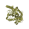
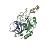
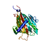
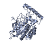
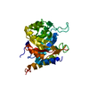
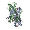
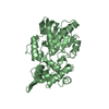
| Title | Crystal structure of the globular domain of human calreticulin | ||||||
 Components Components | calreticulin | ||||||
 Keywords Keywords | CHAPERONE / legume lectin fold / CNX/CRT family / multi-functional / Carbohydrate binding / Peptide Binding / multi-compartmental | ||||||
| Function / homology |  Function and homology information Function and homology informationresponse to biphenyl / Calnexin/calreticulin cycle / negative regulation of intracellular steroid hormone receptor signaling pathway / cytolytic granule / nuclear receptor-mediated glucocorticoid signaling pathway / positive regulation of dendritic cell chemotaxis / Assembly of Viral Components at the Budding Site / ATF6 (ATF6-alpha) activates chaperone genes / cellular response to electrical stimulus / negative regulation of trophoblast cell migration ...response to biphenyl / Calnexin/calreticulin cycle / negative regulation of intracellular steroid hormone receptor signaling pathway / cytolytic granule / nuclear receptor-mediated glucocorticoid signaling pathway / positive regulation of dendritic cell chemotaxis / Assembly of Viral Components at the Budding Site / ATF6 (ATF6-alpha) activates chaperone genes / cellular response to electrical stimulus / negative regulation of trophoblast cell migration / cortical granule / negative regulation of retinoic acid receptor signaling pathway / response to peptide / complement component C1q complex binding / endoplasmic reticulum quality control compartment / protein folding in endoplasmic reticulum / sarcoplasmic reticulum lumen / nuclear export signal receptor activity / cardiac muscle cell differentiation / cellular response to lithium ion / response to glycoside / negative regulation of neuron differentiation / Scavenging by Class A Receptors / Scavenging by Class F Receptors / nuclear androgen receptor binding / response to testosterone / smooth endoplasmic reticulum / Maturation of DENV proteins / hormone binding / molecular sequestering activity / protein localization to nucleus / positive regulation of substrate adhesion-dependent cell spreading / ERAD pathway / endocytic vesicle lumen / protein export from nucleus / peptide binding / endoplasmic reticulum-Golgi intermediate compartment membrane / positive regulation of cell cycle / protein folding chaperone / positive regulation of endothelial cell migration / positive regulation of phagocytosis / acrosomal vesicle / Antigen Presentation: Folding, assembly and peptide loading of class I MHC / lumenal side of endoplasmic reticulum membrane / protein maturation / peptide antigen assembly with MHC class I protein complex / MHC class I peptide loading complex / cellular response to virus / intracellular calcium ion homeostasis / integrin binding / cellular senescence / phagocytic vesicle membrane / : / response to estradiol / nuclear envelope / carbohydrate binding / protein-folding chaperone binding / extracellular matrix / ER-Phagosome pathway / protein folding / spermatogenesis / regulation of apoptotic process / postsynapse / negative regulation of translation / protein stabilization / response to xenobiotic stimulus / ribosome / iron ion binding / endoplasmic reticulum lumen / external side of plasma membrane / focal adhesion / negative regulation of DNA-templated transcription / mRNA binding / positive regulation of gene expression / positive regulation of cell population proliferation / calcium ion binding / ubiquitin protein ligase binding / regulation of DNA-templated transcription / endoplasmic reticulum membrane / perinuclear region of cytoplasm / glutamatergic synapse / negative regulation of transcription by RNA polymerase II / cell surface / endoplasmic reticulum / mitochondrion / DNA binding / : / RNA binding / extracellular exosome / extracellular region / zinc ion binding / membrane / nucleus / cytosol / cytoplasm Similarity search - Function | ||||||
| Biological species |  Homo sapiens (human) Homo sapiens (human) | ||||||
| Method |  X-RAY DIFFRACTION / SYNCHROTRON / MOLECULAR REPLACEMENT / Resolution: 1.55 Å X-RAY DIFFRACTION / SYNCHROTRON / MOLECULAR REPLACEMENT / Resolution: 1.55 Å | ||||||
 Authors Authors | Gaboriaud, C. | ||||||
 Citation Citation | Journal: Plos One / Year: 2011 Title: X-ray structure of the human calreticulin globular domain reveals a Peptide-binding area and suggests a multi-molecular mechanism Authors: Chouquet, A. / Paidassi, H. / Ling, W.L. / Frachet, P. / Houen, G. / Arlaud, G.J. / Gaboriaud, C. | ||||||
| History |
|
- Structure visualization
| Structure viewer | Molecule: MolmilJmol/JSmol |
|---|
- Downloads & links
-Download
| PDBx/mmCIF format | 3pow.cif.gz | 121.1 KB | Display | PDBx/mmCIF format |
|---|---|---|---|---|
| PDB format | pdb3pow.ent.gz | 90.9 KB | Display | PDB format |
| PDBx/mmJSON format | 3pow.json.gz | Tree view | PDBx/mmJSON format | |
| Others |  Other downloads Other downloads |
-Validation report
| Arichive directory | https://data.pdbj.org/pub/pdb/validation_reports/po/3powftp://data.pdbj.org/pub/pdb/validation_reports/po/3pow | HTTPS FTP |
|---|
-Related structure data
| Related structure data |  3posSC S: Starting model for refinement C: citing same article ( |
|---|---|
| Similar structure data |


-Links
 PDBj
PDBj


- Assembly
| Deposited unit | 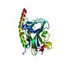
| ||||||||
|---|---|---|---|---|---|---|---|---|---|
| 1 |
| ||||||||
| Unit cell |
|
-Components
| #1: Protein | Mass: 30146.271 Da / Num. of mol.: 1 Fragment: Globular domain, UNP residues 18-204 and 302-368 linked with GSG Source method: isolated from a genetically manipulated source Source: (gene. exp.) Homo sapiens (human) / Gene: CALR, CRTC / Plasmid: pHFX-CRT / Production host:  |
|---|---|
| #2: Chemical | ChemComp-CA /   Mass: 40.078 Da / Num. of mol.: 1 / Source method: obtained synthetically / Formula: Ca Mass: 40.078 Da / Num. of mol.: 1 / Source method: obtained synthetically / Formula: Ca |
| #3: Water | ChemComp-HOH /  Mass: 18.015 Da / Num. of mol.: 334 / Source method: isolated from a natural source / Formula: H2O Mass: 18.015 Da / Num. of mol.: 334 / Source method: isolated from a natural source / Formula: H2O |
| Has protein modification | Y |
-Experimental details
-Experiment
| Experiment | Method: X-RAY DIFFRACTION / Number of used crystals: 1 |
|---|
- Sample preparation
| Crystal | Density Matthews: 2.3 Å3/Da / Density % sol: 46.49 % |
|---|---|
| Crystal grow | Temperature: 277.1 K / Method: vapor diffusion / pH: 6 Details: 30% PEG 4000, 0.2M ammonium acetate, 10mM magnesium acetate, and either 0.05M MES (pH 5.7 or 6.0), or 0.05M HEPES (pH 7.0), VAPOR DIFFUSION, temperature 277.1K |
-Data collection
| Diffraction | Mean temperature: 100 K | ||||||||||||||||||||||||||||||||||||||||||||||||||||||||||||
|---|---|---|---|---|---|---|---|---|---|---|---|---|---|---|---|---|---|---|---|---|---|---|---|---|---|---|---|---|---|---|---|---|---|---|---|---|---|---|---|---|---|---|---|---|---|---|---|---|---|---|---|---|---|---|---|---|---|---|---|---|---|
| Diffraction source | Source: SYNCHROTRON / Site: ESRF  / Beamline: ID23-2 / Wavelength: 0.8726 Å / Beamline: ID23-2 / Wavelength: 0.8726 Å | ||||||||||||||||||||||||||||||||||||||||||||||||||||||||||||
| Detector | Type: MARMOSAIC 225 mm CCD / Detector: CCD / Date: Apr 24, 2010 | ||||||||||||||||||||||||||||||||||||||||||||||||||||||||||||
| Radiation | Monochromator: horizontally side diffracting Silicon 111 crystal Protocol: SINGLE WAVELENGTH / Monochromatic (M) / Laue (L): M / Scattering type: x-ray | ||||||||||||||||||||||||||||||||||||||||||||||||||||||||||||
| Radiation wavelength | Wavelength: 0.8726 Å / Relative weight: 1 | ||||||||||||||||||||||||||||||||||||||||||||||||||||||||||||
| Reflection | Number: 6705 / D res high: 1.54 Å / Num. obs: 6705 / % possible obs: 2.1 | ||||||||||||||||||||||||||||||||||||||||||||||||||||||||||||
| Diffraction reflection shell |
| ||||||||||||||||||||||||||||||||||||||||||||||||||||||||||||
| Reflection | Resolution: 1.54→20 Å / Num. all: 39833 / Num. obs: 39833 / % possible obs: 96.8 % / Observed criterion σ(I): -3 / Redundancy: 4.7 % / Biso Wilson estimate: 20.093 Å2 / Rsym value: 0.07 | ||||||||||||||||||||||||||||||||||||||||||||||||||||||||||||
| Reflection shell |
|
- Processing
| Software |
| ||||||||||||||||||||||||||||||||||||||||||||||||||||||||||||||||||||||||||||||||||||||||||||||||||||||||||||
|---|---|---|---|---|---|---|---|---|---|---|---|---|---|---|---|---|---|---|---|---|---|---|---|---|---|---|---|---|---|---|---|---|---|---|---|---|---|---|---|---|---|---|---|---|---|---|---|---|---|---|---|---|---|---|---|---|---|---|---|---|---|---|---|---|---|---|---|---|---|---|---|---|---|---|---|---|---|---|---|---|---|---|---|---|---|---|---|---|---|---|---|---|---|---|---|---|---|---|---|---|---|---|---|---|---|---|---|---|---|
| Refinement | Method to determine structure: MOLECULAR REPLACEMENT Starting model: PDB ENTRY 3POS Resolution: 1.55→10 Å / Cor.coef. Fo:Fc: 0.9472 / Cor.coef. Fo:Fc free: 0.9465 / Occupancy max: 1 / Occupancy min: 0.5 / Cross valid method: THROUGHOUT / σ(F): 0 / Stereochemistry target values: Engh & Huber
| ||||||||||||||||||||||||||||||||||||||||||||||||||||||||||||||||||||||||||||||||||||||||||||||||||||||||||||
| Displacement parameters | Biso max: 78.04 Å2 / Biso mean: 16.0774 Å2 / Biso min: 5.52 Å2
| ||||||||||||||||||||||||||||||||||||||||||||||||||||||||||||||||||||||||||||||||||||||||||||||||||||||||||||
| Refine analyze | Luzzati coordinate error obs: 0.187 Å | ||||||||||||||||||||||||||||||||||||||||||||||||||||||||||||||||||||||||||||||||||||||||||||||||||||||||||||
| Refinement step | Cycle: LAST / Resolution: 1.55→10 Å
| ||||||||||||||||||||||||||||||||||||||||||||||||||||||||||||||||||||||||||||||||||||||||||||||||||||||||||||
| Refine LS restraints |
| ||||||||||||||||||||||||||||||||||||||||||||||||||||||||||||||||||||||||||||||||||||||||||||||||||||||||||||
| LS refinement shell | Resolution: 1.55→1.59 Å / Total num. of bins used: 20
| ||||||||||||||||||||||||||||||||||||||||||||||||||||||||||||||||||||||||||||||||||||||||||||||||||||||||||||
| Refinement TLS params. | Method: refined / Origin x: 2.1858 Å / Origin y: 10.1515 Å / Origin z: 15.0518 Å
| ||||||||||||||||||||||||||||||||||||||||||||||||||||||||||||||||||||||||||||||||||||||||||||||||||||||||||||
| Refinement TLS group |
|
