 Movie
Movie Controller


+ Open data

- Basic information
| Entry | Database: PDB / ID: 1hia | ||||||
|---|---|---|---|---|---|---|---|
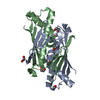
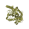
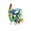
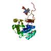
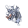
| Title | KALLIKREIN COMPLEXED WITH HIRUSTASIN | ||||||
 Components Components |
| ||||||
 Keywords Keywords | COMPLEX (PROTEASE/INHIBITOR) / COMPLEX (PROTEASE-INHIBITOR) / TISSUE KALLIKREIN / SERINE PROTEASE / TRYPSIN / PSA / KININ / SERPIN / COMPLEX (PROTEASE-INHIBITOR) complex | ||||||
| Function / homology |  Function and homology information Function and homology informationtissue kallikrein / negative regulation of coagulation / regulation of systemic arterial blood pressure / zymogen activation / secretory granule / serine-type endopeptidase inhibitor activity / heparin binding / serine-type endopeptidase activity / : / extracellular region Similarity search - Function | ||||||
| Biological species |   Hirudo medicinalis (medicinal leech) Hirudo medicinalis (medicinal leech) | ||||||
| Method |  X-RAY DIFFRACTION / SYNCHROTRON / MOLECULAR REPLACEMENT / Resolution: 2.4 Å X-RAY DIFFRACTION / SYNCHROTRON / MOLECULAR REPLACEMENT / Resolution: 2.4 Å | ||||||
 Authors Authors | Mittl, P. / Di Marco, S. / Gruetter, M. | ||||||
 Citation Citation | Journal: Structure / Year: 1997 Title: A new structural class of serine protease inhibitors revealed by the structure of the hirustasin-kallikrein complex. Authors: Mittl, P.R. / Di Marco, S. / Fendrich, G. / Pohlig, G. / Heim, J. / Sommerhoff, C. / Fritz, H. / Priestle, J.P. / Grutter, M.G. #1: Journal: Structure / Year: 1997Title: Erratum. A New Structural Class of Serine Protease Inhibitors Revealed by the Structure of the Hirustasin-Kallikrein Complex Authors: Mittl, P.R. / Di Marco, S. / Fendrich, G. / Pohlig, G. / Heim, J. / Sommerhoff, C. / Fritz, H. / Priestle, J.P. / Grutter, M.G. | ||||||
| History |
|
- Structure visualization
| Structure viewer | Molecule: MolmilJmol/JSmol |
|---|
- Downloads & links
-Download
| PDBx/mmCIF format | 1hia.cif.gz | 124.2 KB | Display | PDBx/mmCIF format |
|---|---|---|---|---|
| PDB format | pdb1hia.ent.gz | 97.2 KB | Display | PDB format |
| PDBx/mmJSON format | 1hia.json.gz | Tree view | PDBx/mmJSON format | |
| Others |  Other downloads Other downloads |
-Validation report
| Arichive directory | https://data.pdbj.org/pub/pdb/validation_reports/hi/1hiaftp://data.pdbj.org/pub/pdb/validation_reports/hi/1hia | HTTPS FTP |
|---|
-Related structure data
| Related structure data | 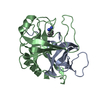 2pkaS S: Starting model for refinement |
|---|---|
| Similar structure data |

-Links
 PDBj
PDBj


- Assembly
| Deposited unit | 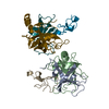
| ||||||||||||
|---|---|---|---|---|---|---|---|---|---|---|---|---|---|
| 1 | 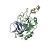
| ||||||||||||
| 2 | 
| ||||||||||||
| Unit cell |
| ||||||||||||
| Noncrystallographic symmetry (NCS) | NCS oper:
|
-Components
| #1: Protein | Mass: 9119.133 Da / Num. of mol.: 2 Source method: isolated from a genetically manipulated source Source: (gene. exp.) #2: Protein | Mass: 16511.477 Da / Num. of mol.: 2 Source method: isolated from a genetically manipulated source Source: (gene. exp.) #3: Protein/peptide | Mass: 5200.117 Da / Num. of mol.: 2 Source method: isolated from a genetically manipulated source Source: (gene. exp.) Hirudo medicinalis (medicinal leech) / Organ: PANCREAS / References: UniProt: P80302#4: Water | ChemComp-HOH / |  Mass: 18.015 Da / Num. of mol.: 304 / Source method: isolated from a natural source / Formula: H2O Mass: 18.015 Da / Num. of mol.: 304 / Source method: isolated from a natural source / Formula: H2OHas protein modification | Y | |
|---|
-Experimental details
-Experiment
| Experiment | Method: X-RAY DIFFRACTION / Number of used crystals: 2 |
|---|
- Sample preparation
| Crystal | Density Matthews: 2.77 Å3/Da / Density % sol: 56.49 % | ||||||||||||||||||||||||||||||||||||||||
|---|---|---|---|---|---|---|---|---|---|---|---|---|---|---|---|---|---|---|---|---|---|---|---|---|---|---|---|---|---|---|---|---|---|---|---|---|---|---|---|---|---|
| Crystal grow | Method: vapor diffusion, hanging drop / pH: 5 Details: HANGING DROP, RESERVOIR: 23% PEG2000, 180MM AM2SO4, 3.5% DIOXANE 100 MM SODIUM ACETATE, PH 4.6. DROP: 1:1 RATIO OF HIRUSTASIN:KALLIKREIN IN 20MM TRIS-HCL, PH 8.0, pH 5.0, vapor diffusion - hanging drop PH range: 4.6-8.0 | ||||||||||||||||||||||||||||||||||||||||
| Crystal grow | *PLUS pH: 8 / Method: vapor diffusion, hanging drop | ||||||||||||||||||||||||||||||||||||||||
| Components of the solutions | *PLUS
|
-Data collection
| Diffraction | Mean temperature: 297 K |
|---|---|
| Diffraction source | Source: SYNCHROTRON / Site: ESRF  / Beamline: BM1A / Wavelength: 0.875 / Beamline: BM1A / Wavelength: 0.875 |
| Detector | Type: MARRESEARCH / Detector: IMAGE PLATE / Date: Nov 1, 1995 |
| Radiation | Monochromator: GRAPHITE(002) / Monochromatic (M) / Laue (L): M / Scattering type: x-ray |
| Radiation wavelength | Wavelength: 0.875 Å / Relative weight: 1 |
| Reflection | Resolution: 2.4→25 Å / Num. obs: 27511 / % possible obs: 97.9 % / Redundancy: 3.5 % / Rsym value: 0.126 / Net I/σ(I): 11.2 |
| Reflection shell | Resolution: 2.4→2.45 Å / Redundancy: 3.1 % / Mean I/σ(I) obs: 2.3 / Rsym value: 0.474 / % possible all: 98.6 |
| Reflection | *PLUS Num. measured all: 96170 / Rmerge(I) obs: 0.126 |
| Reflection shell | *PLUS % possible obs: 98.6 % / Rmerge(I) obs: 0.474 |
- Processing
| Software |
| ||||||||||||||||||||||||||||||||||||||||||||||||||||||||||||
|---|---|---|---|---|---|---|---|---|---|---|---|---|---|---|---|---|---|---|---|---|---|---|---|---|---|---|---|---|---|---|---|---|---|---|---|---|---|---|---|---|---|---|---|---|---|---|---|---|---|---|---|---|---|---|---|---|---|---|---|---|---|
| Refinement | Method to determine structure: MOLECULAR REPLACEMENT Starting model: PDB ENTRY 2PKA Resolution: 2.4→8 Å / Rfactor Rfree error: 0 / Data cutoff high absF: 10000000 / Data cutoff low absF: 0.001 / Cross valid method: THROUGHOUT Details: N-TERMINAL RESIDUES (I/J 5-23) OF HIRUSTASIN HAVE HIGH B-FACTORS. NCS-RELATED MOLECULES ARE RELATED BY THE SAME ORIENTATION (IDENTITY MATRIX) AND THE FRACTIONAL TRANSLATION VECTOR (1/2, 1/3, ...Details: N-TERMINAL RESIDUES (I/J 5-23) OF HIRUSTASIN HAVE HIGH B-FACTORS. NCS-RELATED MOLECULES ARE RELATED BY THE SAME ORIENTATION (IDENTITY MATRIX) AND THE FRACTIONAL TRANSLATION VECTOR (1/2, 1/3, 1/2). THIS PACKING EXERTS THE EXTINCTION PATTERN K=3N: H+L=2N.
| ||||||||||||||||||||||||||||||||||||||||||||||||||||||||||||
| Displacement parameters | Biso mean: 46.2 Å2
| ||||||||||||||||||||||||||||||||||||||||||||||||||||||||||||
| Refinement step | Cycle: LAST / Resolution: 2.4→8 Å
| ||||||||||||||||||||||||||||||||||||||||||||||||||||||||||||
| Refine LS restraints |
| ||||||||||||||||||||||||||||||||||||||||||||||||||||||||||||
| Xplor file |
| ||||||||||||||||||||||||||||||||||||||||||||||||||||||||||||
| Software | *PLUS Name: X-PLOR / Version: 3.1 / Classification: refinement | ||||||||||||||||||||||||||||||||||||||||||||||||||||||||||||
| Refinement | *PLUS | ||||||||||||||||||||||||||||||||||||||||||||||||||||||||||||
| Solvent computation | *PLUS | ||||||||||||||||||||||||||||||||||||||||||||||||||||||||||||
| Displacement parameters | *PLUS | ||||||||||||||||||||||||||||||||||||||||||||||||||||||||||||
| Refine LS restraints | *PLUS
|
