 Movie
Movie Controller


+ Open data

- Basic information
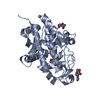
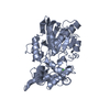
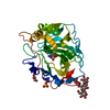
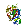
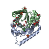
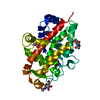
| Entry | Database: PDB / ID: 3ngm | ||||||
|---|---|---|---|---|---|---|---|
| Title | Crystal structure of lipase from Gibberella zeae | ||||||
 Components Components | Extracellular lipase | ||||||
 Keywords Keywords | HYDROLASE / secret lipase / Gibberella zeae | ||||||
| Function / homology |  Function and homology information Function and homology informationdiacylglycerol lipase activity / monoacylglycerol lipase activity / triacylglycerol lipase / triacylglycerol lipase activity / lipid catabolic process Similarity search - Function | ||||||
| Biological species |  Gibberella zeae (fungus) Gibberella zeae (fungus) | ||||||
| Method |  X-RAY DIFFRACTION / MOLECULAR REPLACEMENT / Resolution: 2.8 Å X-RAY DIFFRACTION / MOLECULAR REPLACEMENT / Resolution: 2.8 Å | ||||||
 Authors Authors | Lou, Z.Y. / Li, M. / Sun, Y.N. / Liu, Y. / Liu, Z. / Rao, Z.H. | ||||||
 Citation Citation | Journal: Protein Cell / Year: 2010 Title: Crystal structure of a secreted lipase from Gibberella zeae reveals a novel "double-lock" mechanism Authors: Lou, Z.Y. / Li, M. / Sun, Y.N. / Liu, Y. / Liu, Z. / Wu, W.P. / Rao, Z.H. | ||||||
| History |
|
- Structure visualization
| Structure viewer | Molecule: MolmilJmol/JSmol |
|---|
- Downloads & links
-Download
| PDBx/mmCIF format | 3ngm.cif.gz | 227.5 KB | Display | PDBx/mmCIF format |
|---|---|---|---|---|
| PDB format | pdb3ngm.ent.gz | 184.2 KB | Display | PDB format |
| PDBx/mmJSON format | 3ngm.json.gz | Tree view | PDBx/mmJSON format | |
| Others |  Other downloads Other downloads |
-Validation report
| Arichive directory | https://data.pdbj.org/pub/pdb/validation_reports/ng/3ngmftp://data.pdbj.org/pub/pdb/validation_reports/ng/3ngm | HTTPS FTP |
|---|
-Related structure data
| Related structure data |  1einS S: Starting model for refinement |
|---|---|
| Similar structure data |

-Links
 PDBj
PDBj- Assembly
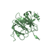
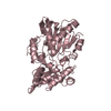
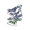
| Deposited unit | 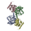
| ||||||||
|---|---|---|---|---|---|---|---|---|---|
| 1 | 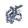
| ||||||||
| 2 | 
| ||||||||
| 3 | 
| ||||||||
| 4 | 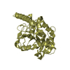
| ||||||||
| Unit cell |
|
-Components
| #1: Protein | Mass: 33799.562 Da / Num. of mol.: 4 / Fragment: residues 1-319 Source method: isolated from a genetically manipulated source Source: (gene. exp.) Gibberella zeae (fungus) / Gene: FGL1 / Plasmid: pLIZG7 / Production host: Pichia pastoris (fungus) / Strain (production host): KM71 / References: UniProt: Q6WER3, triacylglycerol lipase#2: Water | ChemComp-HOH / |  Mass: 18.015 Da / Num. of mol.: 218 / Source method: isolated from a natural source / Formula: H2O Mass: 18.015 Da / Num. of mol.: 218 / Source method: isolated from a natural source / Formula: H2OHas protein modification | Y | |
|---|
-Experimental details
-Experiment
| Experiment | Method: X-RAY DIFFRACTION / Number of used crystals: 1 |
|---|
- Sample preparation
| Crystal | Density Matthews: 2.58 Å3/Da / Density % sol: 52.39 % |
|---|---|
| Crystal grow | Temperature: 291 K / Method: vapor diffusion, hanging drop / pH: 5.5 Details: pH 5.5, VAPOR DIFFUSION, HANGING DROP, temperature 291K |
-Data collection
| Diffraction | Mean temperature: 100 K |
|---|---|
| Diffraction source | Source: ROTATING ANODE / Type: RIGAKU MICROMAX-007 / Wavelength: 1.5418 Å |
| Detector | Type: MAR scanner 345 mm plate / Detector: IMAGE PLATE / Date: Jan 3, 2008 / Details: osmic mirror |
| Radiation | Protocol: SINGLE WAVELENGTH / Monochromatic (M) / Laue (L): M / Scattering type: x-ray |
| Radiation wavelength | Wavelength: 1.5418 Å / Relative weight: 1 |
| Reflection | Resolution: 2.8→50 Å / Num. all: 35284 / Num. obs: 34080 / % possible obs: 98 % / Observed criterion σ(F): 0 / Observed criterion σ(I): 0 / Rmerge(I) obs: 0.101 |
- Processing
| Software |
| ||||||||||||||||||||
|---|---|---|---|---|---|---|---|---|---|---|---|---|---|---|---|---|---|---|---|---|---|
| Refinement | Method to determine structure: MOLECULAR REPLACEMENT Starting model: 1EIN Resolution: 2.8→50 Å / Cross valid method: THROUGHOUT / σ(F): 0 / Stereochemistry target values: Engh & Huber
| ||||||||||||||||||||
| Refinement step | Cycle: LAST / Resolution: 2.8→50 Å
| ||||||||||||||||||||
| Refine LS restraints |
|
