- PDB-5lhw: Central Coiled-Coil Domain of Human STIL -
+
Open data
ID or keywords:
Loading...
-
Basic information
Entry
Database: PDB / ID: 5lhw
Title
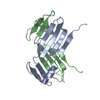
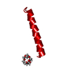
Central Coiled-Coil Domain of Human STIL
Components
SCL-interrupting locus protein
Keywords
STRUCTURAL PROTEIN / Centriole
Function / homology
Function and homology information
floor plate development / regulation of centriole replication / procentriole replication complex / positive regulation of centriole replication / embryonic axis specification / notochord development / microtubule organizing center organization / positive regulation of spindle assembly / protein localization to centrosome / determination of left/right symmetry ...floor plate development / regulation of centriole replication / procentriole replication complex / positive regulation of centriole replication / embryonic axis specification / notochord development / microtubule organizing center organization / positive regulation of spindle assembly / protein localization to centrosome / determination of left/right symmetry / neural tube development / smoothened signaling pathway / heart looping / forebrain development / centrosome duplication / microtubule organizing center / positive regulation of G1/S transition of mitotic cell cycle / mitotic spindle organization / regulation of mitotic spindle organization / neural tube closure / centriole / multicellular organism growth / cell cortex / in utero embryonic development / ciliary basal body / centrosome / negative regulation of apoptotic process / nucleoplasm / identical protein binding / cytoplasm / cytosol Similarity search - Function
: / : / STIL coiled coil region / Proline-rich motif in STIL / SCL-interrupting locus protein / : / STIL N-terminal domain Similarity search - Domain/homology
Mass: 18.015 Da / Num. of mol.: 16 / Source method: isolated from a natural source / Formula: H2O
-
Experimental details
-
Experiment
Experiment
Method: X-RAY DIFFRACTION / Number of used crystals: 1
-
Sample preparation
Crystal
Density Matthews: 1.86 Å3/Da / Density % sol: 33.71 % / Description: Large Block
Crystal grow
Temperature: 292 K / Method: vapor diffusion, sitting drop / pH: 9 Details: 150 nl protein solution (58.6 mg/ml protein in 20 mM Tris pH 7.5, 150 mM NaCl, 2 mM TCEP) and 150 nl of mother liquor (7 mM HEPES pH 8.2, 93 mM Tris pH 9.0, 55.36% v/v PEG 550 MME, 10% v/v glycerol)
In the structure databanks used in Yorodumi, some data are registered as the other names, "COVID-19 virus" and "2019-nCoV". Here are the details of the virus and the list of structure data.
Jan 31, 2019. EMDB accession codes are about to change! (news from PDBe EMDB page)
EMDB accession codes are about to change! (news from PDBe EMDB page)
The allocation of 4 digits for EMDB accession codes will soon come to an end. Whilst these codes will remain in use, new EMDB accession codes will include an additional digit and will expand incrementally as the available range of codes is exhausted. The current 4-digit format prefixed with “EMD-” (i.e. EMD-XXXX) will advance to a 5-digit format (i.e. EMD-XXXXX), and so on. It is currently estimated that the 4-digit codes will be depleted around Spring 2019, at which point the 5-digit format will come into force.
The EM Navigator/Yorodumi systems omit the EMD- prefix.
Related info.:Q: What is EMD? / ID/Accession-code notation in Yorodumi/EM Navigator
Yorodumi is a browser for structure data from EMDB, PDB, SASBDB, etc.
This page is also the successor to EM Navigator detail page, and also detail information page/front-end page for Omokage search.
The word "yorodu" (or yorozu) is an old Japanese word meaning "ten thousand". "mi" (miru) is to see.
Related info.:EMDB / PDB / SASBDB / Comparison of 3 databanks / Yorodumi Search / Aug 31, 2016. New EM Navigator & Yorodumi / Yorodumi Papers / Jmol/JSmol / Function and homology information / Changes in new EM Navigator and Yorodumi
 Movie
Movie Controller


 Open data
 Basic information
 Components
 Keywords
 Function and homology information
 Homo sapiens (human)
 X-RAY DIFFRACTION /
  Authors
 United Kingdom, 2items
  Citation
 Structure visualization
 Downloads & links
 Other downloads


 PDBj
PDBj Assembly
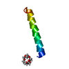


 Mass: 282.331 Da / Num. of mol.: 1 / Source method: obtained synthetically / Formula: C12H26O7 / Comment: precipitant*YM
 Mass: 18.015 Da / Num. of mol.: 16 / Source method: isolated from a natural source / Formula: H2O
 Sample preparation
 / Beamline: ID29 / Wavelength: 0.8 Å
 Processing