 Movie
Movie Controller

[English] 日本語
 Yorodumi
- PDB-5lek: The Transcriptional Regulator PrfA-G145S mutant from Listeria Mon... -
+ Open data

- Basic information
| Entry | Database: PDB / ID: 5lek | |||||||||
|---|---|---|---|---|---|---|---|---|---|---|
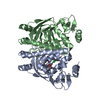
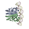
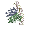
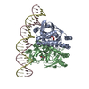
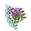
| Title | The Transcriptional Regulator PrfA-G145S mutant from Listeria Monocytogenes in complex with a 30-bp operator PrfA-box motif | |||||||||
 Components Components |
| |||||||||
 Keywords Keywords | TRANSCRIPTION / Transcription regulator / DNA binding / activation / Listeria monocytogenes | |||||||||
| Function / homology |  Function and homology information Function and homology information | |||||||||
| Biological species |  Listeria monocytogenes serovar 1/2a (bacteria)Listeria monocytogenes (bacteria) Listeria monocytogenes serovar 1/2a (bacteria)Listeria monocytogenes (bacteria) | |||||||||
| Method |  X-RAY DIFFRACTION / SYNCHROTRON / MOLECULAR REPLACEMENT / Resolution: 2.8 Å X-RAY DIFFRACTION / SYNCHROTRON / MOLECULAR REPLACEMENT / Resolution: 2.8 Å | |||||||||
 Authors Authors | Hall, M. / Grundstrom, C. / Begum, A. / Lindberg, M. / Sauer, U.H. / Almqvist, F. / Johansson, J. / Sauer-Eriksson, A.E. | |||||||||
| Funding support |  Sweden, 2items Sweden, 2items
| |||||||||
 Citation Citation | Journal: Proc. Natl. Acad. Sci. U.S.A. / Year: 2016 Title: Structural basis for glutathione-mediated activation of the virulence regulatory protein PrfA in Listeria. Authors: Hall, M. / Grundstrom, C. / Begum, A. / Lindberg, M.J. / Sauer, U.H. / Almqvist, F. / Johansson, J. / Sauer-Eriksson, A.E. | |||||||||
| History |
|
- Structure visualization
| Structure viewer | Molecule: MolmilJmol/JSmol |
|---|
- Downloads & links
-Download
| PDBx/mmCIF format | 5lek.cif.gz | 238.7 KB | Display | PDBx/mmCIF format |
|---|---|---|---|---|
| PDB format | pdb5lek.ent.gz | 190.1 KB | Display | PDB format |
| PDBx/mmJSON format | 5lek.json.gz | Tree view | PDBx/mmJSON format | |
| Others |  Other downloads Other downloads |
-Validation report
| Arichive directory | https://data.pdbj.org/pub/pdb/validation_reports/le/5lekftp://data.pdbj.org/pub/pdb/validation_reports/le/5lek | HTTPS FTP |
|---|
-Related structure data
| Related structure data |  5lejC  5lrrC  5lrsC 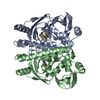 2bgcS S: Starting model for refinement C: citing same article ( |
|---|---|
| Similar structure data |

-Links
 PDBj
PDBj


- Assembly
| Deposited unit | 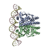
| ||||||||
|---|---|---|---|---|---|---|---|---|---|
| 1 |
| ||||||||
| Unit cell |
|
-Components
| #1: Protein | Mass: 27359.418 Da / Num. of mol.: 2 Source method: isolated from a genetically manipulated source Source: (gene. exp.) Listeria monocytogenes serovar 1/2a (strain ATCC BAA-679 / EGD-e) (bacteria)Gene: prfA, lmo0200 / Production host: #2: DNA chain | | Mass: 9261.000 Da / Num. of mol.: 1 / Source method: obtained synthetically / Source: (synth.) Listeria monocytogenes (bacteria)#3: DNA chain | | Mass: 9180.953 Da / Num. of mol.: 1 / Source method: obtained synthetically / Source: (synth.) Listeria monocytogenes (bacteria)#4: Water | ChemComp-HOH / |  Mass: 18.015 Da / Num. of mol.: 3 / Source method: isolated from a natural source / Formula: H2O Mass: 18.015 Da / Num. of mol.: 3 / Source method: isolated from a natural source / Formula: H2O |
|---|
-Experimental details
-Experiment
| Experiment | Method: X-RAY DIFFRACTION / Number of used crystals: 1 |
|---|
- Sample preparation
| Crystal | Density Matthews: 2.84 Å3/Da / Density % sol: 56.72 % |
|---|---|
| Crystal grow | Temperature: 291 K / Method: vapor diffusion, hanging drop / pH: 5.5 Details: Protein and duplex DNA were incubated together at a ratio of 1:1.3 (PrfA dimer:hly DNA) at final concentrations of 50 microM and 70 microM respectively in 20 mM Tris-HCl pH 8.0, 150 mM NaCl, ...Details: Protein and duplex DNA were incubated together at a ratio of 1:1.3 (PrfA dimer:hly DNA) at final concentrations of 50 microM and 70 microM respectively in 20 mM Tris-HCl pH 8.0, 150 mM NaCl, 1 mM DTT for 60 min at room temperature, before being used for crystal setups. Crystals were obtained after 24 h by mixing 4 microL protein-DNA solution with 2 microL reservoir solution consisting of 8% PEG 8000, 100 mM sodium acetate pH 4.6, 100 mM magnesium acetate, 20% glycerol. Prior to vitrification the soaking of PrfAG145S-DNA crystals were soaked in a reservoir solution containing 30% glycerol for 24 h |
-Data collection
| Diffraction | Mean temperature: 100 K |
|---|---|
| Diffraction source | Source: SYNCHROTRON / Site: BESSY  / Beamline: 14.1 / Wavelength: 0.8729 Å / Beamline: 14.1 / Wavelength: 0.8729 Å |
| Detector | Type: DECTRIS PILATUS 6M-F / Detector: PIXEL / Date: Jan 22, 2016 |
| Radiation | Protocol: SINGLE WAVELENGTH / Monochromatic (M) / Laue (L): M / Scattering type: x-ray |
| Radiation wavelength | Wavelength: 0.8729 Å / Relative weight: 1 |
| Reflection | Resolution: 2.8→47.3 Å / Num. obs: 21616 / % possible obs: 99.4 % / Redundancy: 7 % / Rmerge(I) obs: 0.091 / Net I/σ(I): 14 |
| Reflection shell | Resolution: 2.8→2.95 Å / Redundancy: 6.9 % / Rmerge(I) obs: 1.281 / Mean I/σ(I) obs: 1.7 / % possible all: 99.2 |
- Processing
| Software |
| |||||||||||||||||||||||||||||||||||||||||||||||||||||||||||||||
|---|---|---|---|---|---|---|---|---|---|---|---|---|---|---|---|---|---|---|---|---|---|---|---|---|---|---|---|---|---|---|---|---|---|---|---|---|---|---|---|---|---|---|---|---|---|---|---|---|---|---|---|---|---|---|---|---|---|---|---|---|---|---|---|---|
| Refinement | Method to determine structure: MOLECULAR REPLACEMENT Starting model: 2bgc Resolution: 2.8→47.297 Å / SU ML: 0.5 / Cross valid method: FREE R-VALUE / σ(F): 1.34 / Phase error: 30.24
| |||||||||||||||||||||||||||||||||||||||||||||||||||||||||||||||
| Solvent computation | Shrinkage radii: 0.9 Å / VDW probe radii: 1.11 Å | |||||||||||||||||||||||||||||||||||||||||||||||||||||||||||||||
| Refinement step | Cycle: LAST / Resolution: 2.8→47.297 Å
| |||||||||||||||||||||||||||||||||||||||||||||||||||||||||||||||
| Refine LS restraints |
| |||||||||||||||||||||||||||||||||||||||||||||||||||||||||||||||
| LS refinement shell |
|
