 Movie
Movie Controller

[English] 日本語
 Yorodumi
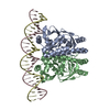
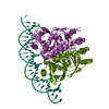
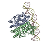
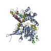
- PDB-6qwf: The Transcriptional Regulator PrfA-A94V mutant from Listeria Mono... -
+ Open data

- Basic information
| Entry | Database: PDB / ID: 6qwf | ||||||
|---|---|---|---|---|---|---|---|
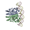
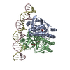
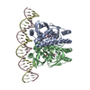
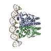
| Title | The Transcriptional Regulator PrfA-A94V mutant from Listeria Monocytogenes in complex with a 30-bp operator PrfA-box motif | ||||||
 Components Components |
| ||||||
 Keywords Keywords | DNA BINDING PROTEIN / TRANSCRIPTION REGULATOR / ACTIVATION / LISTERIA MONOCYTOGENES / TRANSCRIPTION / VIRULENCE FACTOR | ||||||
| Function / homology |  Function and homology information Function and homology informationpositive regulation of single-species biofilm formation on inanimate substrate / DNA-binding transcription factor activity / DNA binding / cytosol Similarity search - Function | ||||||
| Biological species |  Listeria monocytogenes (bacteria) Listeria monocytogenes (bacteria) | ||||||
| Method |  X-RAY DIFFRACTION / SYNCHROTRON / MOLECULAR REPLACEMENT / Resolution: 2.7 Å X-RAY DIFFRACTION / SYNCHROTRON / MOLECULAR REPLACEMENT / Resolution: 2.7 Å | ||||||
 Authors Authors | Hall, M. / Grundstrom, C. / Hansen, S. / Brannstrom, K. / Johansson, J. / Sauer-Eriksson, A.E. | ||||||
| Funding support |  Sweden, 1items Sweden, 1items
| ||||||
 Citation Citation | Journal: J.Bacteriol. / Year: 2020 Title: A Novel Growth-Based Selection Strategy Identifies New Constitutively Active Variants of the Major Virulence Regulator PrfA in Listeria monocytogenes. Authors: Hansen, S. / Hall, M. / Grundstrom, C. / Brannstrom, K. / Sauer-Eriksson, A.E. / Johansson, J. | ||||||
| History |
|
- Structure visualization
| Structure viewer | Molecule: MolmilJmol/JSmol |
|---|
- Downloads & links
-Download
| PDBx/mmCIF format | 6qwf.cif.gz | 143.7 KB | Display | PDBx/mmCIF format |
|---|---|---|---|---|
| PDB format | pdb6qwf.ent.gz | 108 KB | Display | PDB format |
| PDBx/mmJSON format | 6qwf.json.gz | Tree view | PDBx/mmJSON format | |
| Others |  Other downloads Other downloads |
-Validation report
| Arichive directory | https://data.pdbj.org/pub/pdb/validation_reports/qw/6qwfftp://data.pdbj.org/pub/pdb/validation_reports/qw/6qwf | HTTPS FTP |
|---|
-Related structure data
| Related structure data |  6qvyC  6qvzC  6qw1C  6qw2C  6qwhC  6qwkC  6qwmC  5lejS S: Starting model for refinement C: citing same article ( |
|---|---|
| Similar structure data |
-Links
 PDBj
PDBj


- Assembly
| Deposited unit | 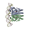
| ||||||||
|---|---|---|---|---|---|---|---|---|---|
| 1 |
| ||||||||
| Unit cell |
|
-Components
| #1: Protein | Mass: 27485.576 Da / Num. of mol.: 2 Source method: isolated from a genetically manipulated source Source: (gene. exp.) Listeria monocytogenes (bacteria) / Gene: prfA, CDR86_15285, D3B95_15085, DOE67_09655 / Production host: #2: DNA chain | | Mass: 9261.000 Da / Num. of mol.: 1 / Source method: obtained synthetically / Source: (synth.) Listeria monocytogenes (bacteria)#3: DNA chain | | Mass: 9180.953 Da / Num. of mol.: 1 / Source method: obtained synthetically / Source: (synth.) Listeria monocytogenes (bacteria)#4: Chemical |   Mass: 92.094 Da / Num. of mol.: 2 / Source method: obtained synthetically / Formula: C3H8O3 Mass: 92.094 Da / Num. of mol.: 2 / Source method: obtained synthetically / Formula: C3H8O3#5: Water | ChemComp-HOH / |  Mass: 18.015 Da / Num. of mol.: 24 / Source method: isolated from a natural source / Formula: H2O Mass: 18.015 Da / Num. of mol.: 24 / Source method: isolated from a natural source / Formula: H2O |
|---|
-Experimental details
-Experiment
| Experiment | Method: X-RAY DIFFRACTION / Number of used crystals: 1 |
|---|
- Sample preparation
| Crystal | Density Matthews: 2.78 Å3/Da / Density % sol: 55.77 % |
|---|---|
| Crystal grow | Temperature: 291 K / Method: vapor diffusion, hanging drop / pH: 8 Details: Droplets of 4 microl protein-DNA solution (ratio 1:1.3) in 20 mM Tris-HCl pH 8.0, 50 mM NaCl, and 1 mM EDTA at 6 mg per ml were mixed with 2 microl reservoir solution consisting of 8% PEG ...Details: Droplets of 4 microl protein-DNA solution (ratio 1:1.3) in 20 mM Tris-HCl pH 8.0, 50 mM NaCl, and 1 mM EDTA at 6 mg per ml were mixed with 2 microl reservoir solution consisting of 8% PEG 8000, 100 mM sodium citrate pH 4.6, 100 mM Magnesium acetate and 20% glycerol. |
-Data collection
| Diffraction | Mean temperature: 100 K / Serial crystal experiment: N |
|---|---|
| Diffraction source | Source: SYNCHROTRON / Site: ESRF  / Beamline: ID30B / Wavelength: 0.9677 Å / Beamline: ID30B / Wavelength: 0.9677 Å |
| Detector | Type: DECTRIS PILATUS3 6M / Detector: PIXEL / Date: Mar 3, 2017 |
| Radiation | Protocol: SINGLE WAVELENGTH / Monochromatic (M) / Laue (L): M / Scattering type: x-ray |
| Radiation wavelength | Wavelength: 0.9677 Å / Relative weight: 1 |
| Reflection | Resolution: 2.7→42.56 Å / Num. obs: 23582 / % possible obs: 99.9 % / Redundancy: 26.8 % / Rmerge(I) obs: 0.097 / Rpim(I) all: 0.026 / Net I/σ(I): 21.2 |
| Reflection shell | Resolution: 2.7→2.8 Å / Redundancy: 28.3 % / Rmerge(I) obs: 1.367 / Mean I/σ(I) obs: 3.2 / Num. unique obs: 2304 / Rpim(I) all: 0.363 / % possible all: 100 |
- Processing
| Software |
| |||||||||||||||||||||||||||||||||||||||||||||||||||||||||||||||
|---|---|---|---|---|---|---|---|---|---|---|---|---|---|---|---|---|---|---|---|---|---|---|---|---|---|---|---|---|---|---|---|---|---|---|---|---|---|---|---|---|---|---|---|---|---|---|---|---|---|---|---|---|---|---|---|---|---|---|---|---|---|---|---|---|
| Refinement | Method to determine structure: MOLECULAR REPLACEMENT Starting model: 5LEJ Resolution: 2.7→42.557 Å / SU ML: 0.4 / Cross valid method: FREE R-VALUE / σ(F): 1.34 / Phase error: 30.12
| |||||||||||||||||||||||||||||||||||||||||||||||||||||||||||||||
| Solvent computation | Shrinkage radii: 0.9 Å / VDW probe radii: 1.11 Å | |||||||||||||||||||||||||||||||||||||||||||||||||||||||||||||||
| Refinement step | Cycle: LAST / Resolution: 2.7→42.557 Å
| |||||||||||||||||||||||||||||||||||||||||||||||||||||||||||||||
| Refine LS restraints |
| |||||||||||||||||||||||||||||||||||||||||||||||||||||||||||||||
| LS refinement shell |
|
