 Movie
Movie Controller

[English] 日本語
 Yorodumi
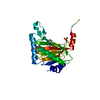
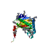
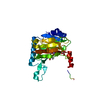
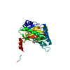
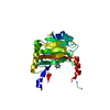
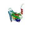
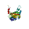
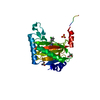
- PDB-5lat: HIF prolyl hydroxylase 2 (PHD2/EGLN1) P317R variant in complex wi... -
+ Open data

- Basic information
| Entry | Database: PDB / ID: 5lat | ||||||
|---|---|---|---|---|---|---|---|
| Title | HIF prolyl hydroxylase 2 (PHD2/EGLN1) P317R variant in complex with Mn(II) and N-[(1-chloro-4-hydroxyisoquinolin-3-yl)carbonyl]glycine (IOX3/UN9) | ||||||
 Components Components | Egl nine homolog 1 | ||||||
 Keywords Keywords | OXIDOREDUCTASE / NON-HEME DIOXYGENASE / IRON / 2-OXOGLUTARATE / HYPOXIA-INDUCIBLE FACTOR / HIF / HIF PROLYL HYDROXYLASE DOMAIN 2 / PHD2 / EGLN1 / OXYGENASE / HYPOXIA / DNA-BINDING / METAL-BINDING / TRANSCRIPTION / HELIX-LOOP-HELIX-BETA / DSBH / FACIAL TRIAD / CYTOPLASM / TRANSCRIPTION/EPIGENETIC REGULATION / SIGNALING / DEVELOPMENT / CELL STRUCTURE / BETA-HYDROXYLATION / TRANSCRIPTION ACTIVATOR/INHIBITOR / UBL CONJUGATION / POLYMORPHISM / VITAMIN C / ZINC-FINGER / FAMILIAL ERYTHROCYTOSIS / BREAST CANCER / TRANSCRIPTION COMPLEX | ||||||
| Function / homology |  Function and homology information Function and homology informationpeptidyl-proline 4-dioxygenase activity / hypoxia-inducible factor-proline dioxygenase / hypoxia-inducible factor-proline dioxygenase activity / negative regulation of hypoxia-inducible factor-1alpha signaling pathway / peptidyl-proline dioxygenase activity / regulation protein catabolic process at postsynapse / intracellular oxygen homeostasis / labyrinthine layer development / cardiac muscle tissue morphogenesis / 2-oxoglutarate-dependent dioxygenase activity ...peptidyl-proline 4-dioxygenase activity / hypoxia-inducible factor-proline dioxygenase / hypoxia-inducible factor-proline dioxygenase activity / negative regulation of hypoxia-inducible factor-1alpha signaling pathway / peptidyl-proline dioxygenase activity / regulation protein catabolic process at postsynapse / intracellular oxygen homeostasis / labyrinthine layer development / cardiac muscle tissue morphogenesis / 2-oxoglutarate-dependent dioxygenase activity / heart trabecula formation / regulation of modification of postsynaptic structure / L-ascorbic acid binding / response to nitric oxide / ventricular septum morphogenesis / regulation of angiogenesis / enzyme inhibitor activity / regulation of neuron apoptotic process / ferrous iron binding / Oxygen-dependent proline hydroxylation of Hypoxia-inducible Factor Alpha / cellular response to hypoxia / intracellular iron ion homeostasis / response to hypoxia / postsynaptic density / glutamatergic synapse / enzyme binding / positive regulation of transcription by RNA polymerase II / zinc ion binding / nucleus / cytoplasm / cytosol Similarity search - Function | ||||||
| Biological species |  Homo sapiens (human) Homo sapiens (human) | ||||||
| Method |  X-RAY DIFFRACTION / SYNCHROTRON / MOLECULAR REPLACEMENT / Resolution: 1.9 Å X-RAY DIFFRACTION / SYNCHROTRON / MOLECULAR REPLACEMENT / Resolution: 1.9 Å | ||||||
 Authors Authors | Chowdhury, R. / Schofield, C.J. | ||||||
 Citation Citation | Journal: Nat Commun / Year: 2016 Title: Structural basis for oxygen degradation domain selectivity of the HIF prolyl hydroxylases. Authors: Chowdhury, R. / Leung, I.K. / Tian, Y.M. / Abboud, M.I. / Ge, W. / Domene, C. / Cantrelle, F.X. / Landrieu, I. / Hardy, A.P. / Pugh, C.W. / Ratcliffe, P.J. / Claridge, T.D. / Schofield, C.J. #1: Journal: ACS Chem. Biol. / Year: 2013Title: Selective small molecule probes for the hypoxia inducible factor (HIF) prolyl hydroxylases. Authors: Chowdhury, R. / Candela-Lena, J.I. / Chan, M.C. / Greenald, D.J. / Yeoh, K.K. / Tian, Y.M. / McDonough, M.A. / Tumber, A. / Rose, N.R. / Conejo-Garcia, A. / Demetriades, M. / Mathavan, S. / ...Authors: Chowdhury, R. / Candela-Lena, J.I. / Chan, M.C. / Greenald, D.J. / Yeoh, K.K. / Tian, Y.M. / McDonough, M.A. / Tumber, A. / Rose, N.R. / Conejo-Garcia, A. / Demetriades, M. / Mathavan, S. / Kawamura, A. / Lee, M.K. / van Eeden, F. / Pugh, C.W. / Ratcliffe, P.J. / Schofield, C.J. #2: Journal: Structure / Year: 2009Title: Structural basis for binding of hypoxia-inducible factor to the oxygen-sensing prolyl hydroxylases. Authors: Chowdhury, R. / McDonough, M.A. / Mecinovic, J. / Loenarz, C. / Flashman, E. / Hewitson, K.S. / Domene, C. / Schofield, C.J. | ||||||
| History |
|
- Structure visualization
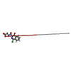
| Structure viewer | Molecule: MolmilJmol/JSmol |
|---|
- Downloads & links
-Download
| PDBx/mmCIF format | 5lat.cif.gz | 149.7 KB | Display | PDBx/mmCIF format |
|---|---|---|---|---|
| PDB format | pdb5lat.ent.gz | 118.4 KB | Display | PDB format |
| PDBx/mmJSON format | 5lat.json.gz | Tree view | PDBx/mmJSON format | |
| Others |  Other downloads Other downloads |
-Validation report
| Arichive directory | https://data.pdbj.org/pub/pdb/validation_reports/la/5latftp://data.pdbj.org/pub/pdb/validation_reports/la/5lat | HTTPS FTP |
|---|
-Related structure data
| Related structure data |  5l9bC  5l9rC  5l9vC 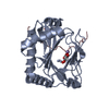 5la9C  5lasC  5lb6C  5lbbC  5lbcC  5lbeC 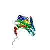 5lbfC 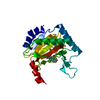 4bqxS S: Starting model for refinement C: citing same article ( |
|---|---|
| Similar structure data |

-Links
 PDBj
PDBj


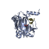
- Assembly
| Deposited unit | 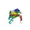
| ||||||||
|---|---|---|---|---|---|---|---|---|---|
| 1 |
| ||||||||
| Unit cell |
|
-Components
-Protein , 1 types, 1 molecules A
| #1: Protein | Mass: 28157.020 Da / Num. of mol.: 1 / Fragment: CATALYTIC DOMAIN, UNP residues 181-426 / Mutation: P317R Source method: isolated from a genetically manipulated source Source: (gene. exp.) Homo sapiens (human) / Gene: EGLN1, C1orf12, PNAS-118, PNAS-137 / Production host:  References: UniProt: Q9GZT9, hypoxia-inducible factor-proline dioxygenase |
|---|
-Non-polymers , 5 types, 190 molecules 


| #2: Chemical | ChemComp-MN /  Mass: 54.938 Da / Num. of mol.: 1 / Source method: obtained synthetically / Formula: Mn Mass: 54.938 Da / Num. of mol.: 1 / Source method: obtained synthetically / Formula: Mn |
|---|---|
| #3: Chemical | ChemComp-UN9 /  Mass: 280.664 Da / Num. of mol.: 1 / Source method: obtained synthetically / Formula: C12H9ClN2O4 Mass: 280.664 Da / Num. of mol.: 1 / Source method: obtained synthetically / Formula: C12H9ClN2O4 |
| #4: Chemical | ChemComp-GOL /  Mass: 92.094 Da / Num. of mol.: 1 / Source method: obtained synthetically / Formula: C3H8O3 Mass: 92.094 Da / Num. of mol.: 1 / Source method: obtained synthetically / Formula: C3H8O3 |
| #5: Chemical | ChemComp-BCT /  Mass: 61.017 Da / Num. of mol.: 1 / Source method: obtained synthetically / Formula: CHO3 / Comment: pH buffer*YM Mass: 61.017 Da / Num. of mol.: 1 / Source method: obtained synthetically / Formula: CHO3 / Comment: pH buffer*YM |
| #6: Water | ChemComp-HOH / Mass: 18.015 Da / Num. of mol.: 186 / Source method: isolated from a natural source / Formula: H2O |
-Details
| Has protein modification | Y |
|---|
-Experimental details
-Experiment
| Experiment | Method: X-RAY DIFFRACTION / Number of used crystals: 1 |
|---|
- Sample preparation
| Crystal | Density Matthews: 2.87 Å3/Da / Density % sol: 57.19 % |
|---|---|
| Crystal grow | Temperature: 293 K / Method: vapor diffusion, sitting drop / pH: 5.6 Details: 0.1 M tri-sodium citrate pH 5.6, 20% PEG 4000, 20% 2-propanol |
-Data collection
| Diffraction | Mean temperature: 100 K |
|---|---|
| Diffraction source | Source: SYNCHROTRON / Site: Diamond  / Beamline: I04 / Wavelength: 0.9795 Å / Beamline: I04 / Wavelength: 0.9795 Å |
| Detector | Type: ADSC QUANTUM 315 / Detector: CCD / Date: Nov 29, 2007 / Details: MIRRORS |
| Radiation | Monochromator: SI 111 / Protocol: SINGLE WAVELENGTH / Monochromatic (M) / Laue (L): M / Scattering type: x-ray |
| Radiation wavelength | Wavelength: 0.9795 Å / Relative weight: 1 |
| Reflection | Resolution: 1.9→27.8 Å / Num. obs: 22463 / % possible obs: 99.6 % / Redundancy: 2.8 % / Biso Wilson estimate: 21.36 Å2 / CC1/2: 0.991 / Rmerge(I) obs: 0.088 / Net I/σ(I): 9.1 |
| Reflection shell | Resolution: 1.9→2 Å / Redundancy: 2.9 % / Rmerge(I) obs: 0.354 / Mean I/σ(I) obs: 2.9 / % possible all: 99.8 |
- Processing
| Software |
| |||||||||||||||||||||||||||||||||||||||||||||||||||||||||||||||||||||||||||||||||||||||||||||||||||||||||||||||||||||||||||||||||||||||||||||||||||||||||||||||||||||||||||||||||||||||||||||||||||||||||||||||||||||||||||||||||||||||||||||||||||||||||||||||||||||||||||||||||||||||||||||||||||||||||||||||||||||||||||||||||||||||||||||||||||||||||||||||||||||||||||||||||||||||||||||||||||||||||||||||||||||||||||||||||||||||||
|---|---|---|---|---|---|---|---|---|---|---|---|---|---|---|---|---|---|---|---|---|---|---|---|---|---|---|---|---|---|---|---|---|---|---|---|---|---|---|---|---|---|---|---|---|---|---|---|---|---|---|---|---|---|---|---|---|---|---|---|---|---|---|---|---|---|---|---|---|---|---|---|---|---|---|---|---|---|---|---|---|---|---|---|---|---|---|---|---|---|---|---|---|---|---|---|---|---|---|---|---|---|---|---|---|---|---|---|---|---|---|---|---|---|---|---|---|---|---|---|---|---|---|---|---|---|---|---|---|---|---|---|---|---|---|---|---|---|---|---|---|---|---|---|---|---|---|---|---|---|---|---|---|---|---|---|---|---|---|---|---|---|---|---|---|---|---|---|---|---|---|---|---|---|---|---|---|---|---|---|---|---|---|---|---|---|---|---|---|---|---|---|---|---|---|---|---|---|---|---|---|---|---|---|---|---|---|---|---|---|---|---|---|---|---|---|---|---|---|---|---|---|---|---|---|---|---|---|---|---|---|---|---|---|---|---|---|---|---|---|---|---|---|---|---|---|---|---|---|---|---|---|---|---|---|---|---|---|---|---|---|---|---|---|---|---|---|---|---|---|---|---|---|---|---|---|---|---|---|---|---|---|---|---|---|---|---|---|---|---|---|---|---|---|---|---|---|---|---|---|---|---|---|---|---|---|---|---|---|---|---|---|---|---|---|---|---|---|---|---|---|---|---|---|---|---|---|---|---|---|---|---|---|---|---|---|---|---|---|---|---|---|---|---|---|---|---|---|---|---|---|---|---|---|---|---|---|---|---|---|---|---|---|---|---|---|---|---|---|---|---|---|---|---|---|---|---|---|---|---|---|---|---|---|---|---|---|---|---|---|---|---|---|---|---|---|---|---|---|---|---|---|---|---|---|---|---|---|---|---|---|---|---|---|---|---|---|---|---|---|---|---|---|---|---|---|---|
| Refinement | Method to determine structure: MOLECULAR REPLACEMENT Starting model: 4BQX Resolution: 1.9→27.796 Å / SU ML: 0.16 / Cross valid method: THROUGHOUT / σ(F): 1.35 / Phase error: 16.09 / Stereochemistry target values: ML
| |||||||||||||||||||||||||||||||||||||||||||||||||||||||||||||||||||||||||||||||||||||||||||||||||||||||||||||||||||||||||||||||||||||||||||||||||||||||||||||||||||||||||||||||||||||||||||||||||||||||||||||||||||||||||||||||||||||||||||||||||||||||||||||||||||||||||||||||||||||||||||||||||||||||||||||||||||||||||||||||||||||||||||||||||||||||||||||||||||||||||||||||||||||||||||||||||||||||||||||||||||||||||||||||||||||||||
| Solvent computation | Shrinkage radii: 0.9 Å / VDW probe radii: 1.11 Å / Solvent model: FLAT MODEL / Bsol: 68.8 Å2 / ksol: 0.42 e/Å3 | |||||||||||||||||||||||||||||||||||||||||||||||||||||||||||||||||||||||||||||||||||||||||||||||||||||||||||||||||||||||||||||||||||||||||||||||||||||||||||||||||||||||||||||||||||||||||||||||||||||||||||||||||||||||||||||||||||||||||||||||||||||||||||||||||||||||||||||||||||||||||||||||||||||||||||||||||||||||||||||||||||||||||||||||||||||||||||||||||||||||||||||||||||||||||||||||||||||||||||||||||||||||||||||||||||||||||
| Displacement parameters | Biso mean: 33 Å2
| |||||||||||||||||||||||||||||||||||||||||||||||||||||||||||||||||||||||||||||||||||||||||||||||||||||||||||||||||||||||||||||||||||||||||||||||||||||||||||||||||||||||||||||||||||||||||||||||||||||||||||||||||||||||||||||||||||||||||||||||||||||||||||||||||||||||||||||||||||||||||||||||||||||||||||||||||||||||||||||||||||||||||||||||||||||||||||||||||||||||||||||||||||||||||||||||||||||||||||||||||||||||||||||||||||||||||
| Refine analyze |
| |||||||||||||||||||||||||||||||||||||||||||||||||||||||||||||||||||||||||||||||||||||||||||||||||||||||||||||||||||||||||||||||||||||||||||||||||||||||||||||||||||||||||||||||||||||||||||||||||||||||||||||||||||||||||||||||||||||||||||||||||||||||||||||||||||||||||||||||||||||||||||||||||||||||||||||||||||||||||||||||||||||||||||||||||||||||||||||||||||||||||||||||||||||||||||||||||||||||||||||||||||||||||||||||||||||||||
| Refinement step | Cycle: LAST / Resolution: 1.9→27.796 Å
| |||||||||||||||||||||||||||||||||||||||||||||||||||||||||||||||||||||||||||||||||||||||||||||||||||||||||||||||||||||||||||||||||||||||||||||||||||||||||||||||||||||||||||||||||||||||||||||||||||||||||||||||||||||||||||||||||||||||||||||||||||||||||||||||||||||||||||||||||||||||||||||||||||||||||||||||||||||||||||||||||||||||||||||||||||||||||||||||||||||||||||||||||||||||||||||||||||||||||||||||||||||||||||||||||||||||||
| Refine LS restraints |
| |||||||||||||||||||||||||||||||||||||||||||||||||||||||||||||||||||||||||||||||||||||||||||||||||||||||||||||||||||||||||||||||||||||||||||||||||||||||||||||||||||||||||||||||||||||||||||||||||||||||||||||||||||||||||||||||||||||||||||||||||||||||||||||||||||||||||||||||||||||||||||||||||||||||||||||||||||||||||||||||||||||||||||||||||||||||||||||||||||||||||||||||||||||||||||||||||||||||||||||||||||||||||||||||||||||||||
| LS refinement shell |
| |||||||||||||||||||||||||||||||||||||||||||||||||||||||||||||||||||||||||||||||||||||||||||||||||||||||||||||||||||||||||||||||||||||||||||||||||||||||||||||||||||||||||||||||||||||||||||||||||||||||||||||||||||||||||||||||||||||||||||||||||||||||||||||||||||||||||||||||||||||||||||||||||||||||||||||||||||||||||||||||||||||||||||||||||||||||||||||||||||||||||||||||||||||||||||||||||||||||||||||||||||||||||||||||||||||||||
| Refinement TLS params. | Method: refined / Refine-ID: X-RAY DIFFRACTION
| |||||||||||||||||||||||||||||||||||||||||||||||||||||||||||||||||||||||||||||||||||||||||||||||||||||||||||||||||||||||||||||||||||||||||||||||||||||||||||||||||||||||||||||||||||||||||||||||||||||||||||||||||||||||||||||||||||||||||||||||||||||||||||||||||||||||||||||||||||||||||||||||||||||||||||||||||||||||||||||||||||||||||||||||||||||||||||||||||||||||||||||||||||||||||||||||||||||||||||||||||||||||||||||||||||||||||
| Refinement TLS group |
|
