 Movie
Movie Controller

[English] 日本語
 Yorodumi
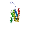
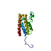
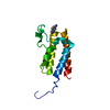
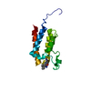
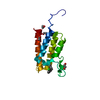
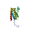
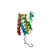
- PDB-5l96: Crystal Structure of BAZ2B bromodomain in complex with 3-amino-2-... -
+ Open data

- Basic information
| Entry | Database: PDB / ID: 5l96 | |||||||||
|---|---|---|---|---|---|---|---|---|---|---|
| Title | Crystal Structure of BAZ2B bromodomain in complex with 3-amino-2-methylpyridine derivative 1 | |||||||||
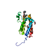
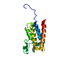
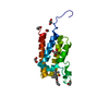
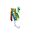
 Components Components | Bromodomain adjacent to zinc finger domain protein 2B | |||||||||
 Keywords Keywords | TRANSCRIPTION / four helical bundle | |||||||||
| Function / homology |  Function and homology information Function and homology informationchromatin remodeling / regulation of transcription by RNA polymerase II / chromatin / DNA binding / zinc ion binding / nucleus Similarity search - Function | |||||||||
| Biological species |  Homo sapiens (human) Homo sapiens (human) | |||||||||
| Method |  X-RAY DIFFRACTION / SYNCHROTRON / MOLECULAR REPLACEMENT / Resolution: 2.15 Å X-RAY DIFFRACTION / SYNCHROTRON / MOLECULAR REPLACEMENT / Resolution: 2.15 Å | |||||||||
 Authors Authors | Lolli, G. / Marchand, J.-R. / Caflisch, A. | |||||||||
| Funding support |  Switzerland, 2items Switzerland, 2items
| |||||||||
 Citation Citation | Journal: J. Med. Chem. / Year: 2016 Title: Derivatives of 3-Amino-2-methylpyridine as BAZ2B Bromodomain Ligands: In Silico Discovery and in Crystallo Validation. Authors: Marchand, J.R. / Lolli, G. / Caflisch, A. | |||||||||
| History |
|
- Structure visualization
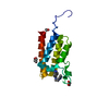
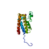
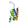
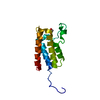
| Structure viewer | Molecule: MolmilJmol/JSmol |
|---|
- Downloads & links
-Download
| PDBx/mmCIF format | 5l96.cif.gz | 64.5 KB | Display | PDBx/mmCIF format |
|---|---|---|---|---|
| PDB format | pdb5l96.ent.gz | 46.2 KB | Display | PDB format |
| PDBx/mmJSON format | 5l96.json.gz | Tree view | PDBx/mmJSON format | |
| Others |  Other downloads Other downloads |
-Validation report
| Arichive directory | https://data.pdbj.org/pub/pdb/validation_reports/l9/5l96ftp://data.pdbj.org/pub/pdb/validation_reports/l9/5l96 | HTTPS FTP |
|---|
-Related structure data
| Related structure data | 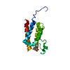 5l8tC 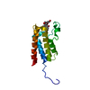 5l8uC  5l97C  5l98C 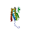 5l99C  4ir5S C: citing same article ( S: Starting model for refinement |
|---|---|
| Similar structure data |


-Links
 PDBj
PDBj
- Assembly
| Deposited unit | 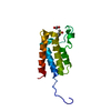
| ||||||||
|---|---|---|---|---|---|---|---|---|---|
| 1 |
| ||||||||
| Unit cell |
|
-Components
| #1: Protein | Mass: 13531.574 Da / Num. of mol.: 1 / Fragment: Bromodomain (residues 2054-2168) Mutation: First two residues SM derive from the expression tag Source method: isolated from a genetically manipulated source Source: (gene. exp.) Homo sapiens (human) / Gene: BAZ2B, KIAA1476 / Production host:  |
|---|---|
| #2: Chemical | ChemComp-6RZ / 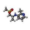  Mass: 228.311 Da / Num. of mol.: 1 / Source method: obtained synthetically / Formula: C10H16N2O2S Mass: 228.311 Da / Num. of mol.: 1 / Source method: obtained synthetically / Formula: C10H16N2O2S |
| #3: Chemical | ChemComp-EDO /   Mass: 62.068 Da / Num. of mol.: 1 / Source method: obtained synthetically / Formula: C2H6O2 Mass: 62.068 Da / Num. of mol.: 1 / Source method: obtained synthetically / Formula: C2H6O2 |
| #4: Water | ChemComp-HOH /  Mass: 18.015 Da / Num. of mol.: 66 / Source method: isolated from a natural source / Formula: H2O Mass: 18.015 Da / Num. of mol.: 66 / Source method: isolated from a natural source / Formula: H2O |
-Experimental details
-Experiment
| Experiment | Method: X-RAY DIFFRACTION / Number of used crystals: 1 |
|---|
- Sample preparation
| Crystal | Density Matthews: 4.24 Å3/Da / Density % sol: 70.99 % / Mosaicity: 0.45 ° |
|---|---|
| Crystal grow | Temperature: 277 K / Method: vapor diffusion, sitting drop / pH: 7.5 Details: PEG500MME (20%), PEG1000 (2%), PEG3350 (2%), PEG20000 (10%), MPD (2%) |
-Data collection
| Diffraction | Mean temperature: 100 K | ||||||||||||||||||||||||||||||
|---|---|---|---|---|---|---|---|---|---|---|---|---|---|---|---|---|---|---|---|---|---|---|---|---|---|---|---|---|---|---|---|
| Diffraction source | Source: SYNCHROTRON / Site: ELETTRA  / Beamline: 5.2R / Wavelength: 1 Å / Beamline: 5.2R / Wavelength: 1 Å | ||||||||||||||||||||||||||||||
| Detector | Type: DECTRIS PILATUS 2M / Detector: PIXEL / Date: Feb 20, 2016 | ||||||||||||||||||||||||||||||
| Radiation | Protocol: SINGLE WAVELENGTH / Monochromatic (M) / Laue (L): M / Scattering type: x-ray | ||||||||||||||||||||||||||||||
| Radiation wavelength | Wavelength: 1 Å / Relative weight: 1 | ||||||||||||||||||||||||||||||
| Reflection | Resolution: 2.15→42.48 Å / Num. obs: 12723 / % possible obs: 99.3 % / Redundancy: 5.1 % / Biso Wilson estimate: 36.08 Å2 / CC1/2: 0.999 / Rmerge(I) obs: 0.069 / Net I/σ(I): 13 | ||||||||||||||||||||||||||||||
| Reflection shell | Diffraction-ID: 1 / Rejects: _
|
- Processing
| Software |
| |||||||||||||||||||||||||||||||||||
|---|---|---|---|---|---|---|---|---|---|---|---|---|---|---|---|---|---|---|---|---|---|---|---|---|---|---|---|---|---|---|---|---|---|---|---|---|
| Refinement | Method to determine structure: MOLECULAR REPLACEMENT Starting model: 4IR5 Resolution: 2.15→33.429 Å / SU ML: 0.19 / Cross valid method: FREE R-VALUE / σ(F): 1.34 / Phase error: 29.34
| |||||||||||||||||||||||||||||||||||
| Solvent computation | Shrinkage radii: 0.9 Å / VDW probe radii: 1.11 Å | |||||||||||||||||||||||||||||||||||
| Displacement parameters | Biso max: 117.14 Å2 / Biso mean: 54.563 Å2 / Biso min: 32.13 Å2 | |||||||||||||||||||||||||||||||||||
| Refinement step | Cycle: final / Resolution: 2.15→33.429 Å
| |||||||||||||||||||||||||||||||||||
| Refine LS restraints |
| |||||||||||||||||||||||||||||||||||
| LS refinement shell | Refine-ID: X-RAY DIFFRACTION / Total num. of bins used: 4
|
