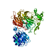
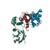
Entry Database : PDB / ID : 5l6wTitle Structure Of the LIMK1-ATPgammaS-CFL1 Complex Cofilin-1 LIM domain kinase 1 Keywords / Function / homology Function Domain/homology Component
/ / / / / / / / / / / / / / / / / / / / / / / / / / / / / / / / / / / / / / / / / / / / / / / / / / / / / / / / / / / / / / / / / / / / / / / / / / / / / / / / / / / / / / / / / / / / / / / / / / / / / / / / / / / / / / / / / / / / / / / / / / / / / / / / / / / / / / / / / / / / / / / / / / / / / / / Biological species Homo sapiens (human)Method / / / Resolution : 2.53 Å Authors Salah, E. / Mathea, S. / Oerum, S. / Newman, J.A. / Tallant, C. / Adamson, R. / Canning, P. / Beltrami, A. / von Delft, F. / Arrowsmith, C.H. ...Salah, E. / Mathea, S. / Oerum, S. / Newman, J.A. / Tallant, C. / Adamson, R. / Canning, P. / Beltrami, A. / von Delft, F. / Arrowsmith, C.H. / Edwards, A.M. / Bountra, C. / Knapp, S. / Bullock, A.N. Journal : To Be Published Title : Structure Of the LIMK1-ATPgammaS-CFL1 ComplexAuthors : Salah, E. / Bullock, A.N. History Deposition May 31, 2016 Deposition site / Processing site Revision 1.0 Jun 8, 2016 Provider / Type Revision 1.1 Oct 16, 2019 Group / Category Revision 1.2 Jan 10, 2024 Group / Database references / Refinement descriptionCategory chem_comp_atom / chem_comp_bond ... chem_comp_atom / chem_comp_bond / database_2 / pdbx_initial_refinement_model Item / _database_2.pdbx_database_accession
Show all Show less
 Movie
Movie Controller


 Open data
 Basic information
 Components
 Keywords
 Function and homology information
 Homo sapiens (human)
 X-RAY DIFFRACTION /
  Authors
 Citation
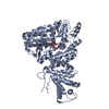
 Structure visualization
 Downloads & links
 Other downloads
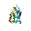


 PDBj
PDBj


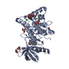
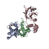
 Assembly
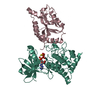

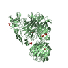
 Spodoptera frugiperda (fall armyworm)

 Mass: 523.247 Da / Num. of mol.: 1 / Source method: obtained synthetically / Formula: C10H16N5O12P3S / Comment: ATP-gamma-S, energy-carrying molecule analogue*YM
 Sample preparation
 / Beamline: I02 / Wavelength: 0.97949 Å
 Processing