 Movie
Movie Controller

[English] 日本語
 Yorodumi
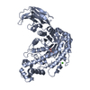
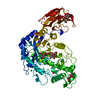
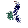
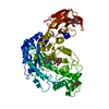
- PDB-5hvk: Crystal structure of LIMK1 mutant D460N in complex with full-leng... -
+ Open data

- Basic information
| Entry | Database: PDB / ID: 5hvk | |||||||||
|---|---|---|---|---|---|---|---|---|---|---|
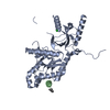
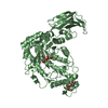
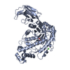
| Title | Crystal structure of LIMK1 mutant D460N in complex with full-length cofilin-1 | |||||||||
 Components Components |
| |||||||||
 Keywords Keywords | TRANSFERASE / kinase substrate actin-remodeling | |||||||||
| Function / homology |  Function and homology information Function and homology informationcellular response to ether / cofilin-actin rod / positive regulation of protein localization to cell leading edge / positive regulation of establishment of cell polarity regulating cell shape / neural fold formation / negative regulation of unidimensional cell growth / positive regulation of barbed-end actin filament capping / negative regulation of lamellipodium assembly / negative regulation of postsynaptic density organization / actin filament fragmentation ...cellular response to ether / cofilin-actin rod / positive regulation of protein localization to cell leading edge / positive regulation of establishment of cell polarity regulating cell shape / neural fold formation / negative regulation of unidimensional cell growth / positive regulation of barbed-end actin filament capping / negative regulation of lamellipodium assembly / negative regulation of postsynaptic density organization / actin filament fragmentation / positive regulation of actin filament depolymerization / negative regulation of actin filament bundle assembly / positive regulation of embryonic development / modification of postsynaptic actin cytoskeleton / positive regulation of actin filament bundle assembly / negative regulation of actin filament depolymerization / actin filament severing / host-mediated activation of viral process / positive regulation of synaptic plasticity / regulation of dendritic spine morphogenesis / establishment of spindle localization / negative regulation of cell adhesion / negative regulation of cell motility / actin filament depolymerization / RHO GTPases Activate ROCKs / axon extension / negative regulation of cell size / cellular response to interleukin-6 / Sema4D induced cell migration and growth-cone collapse / regulation of cell morphogenesis / cell projection organization / negative regulation of dendritic spine maintenance / positive regulation of cell motility / neural crest cell migration / cortical actin cytoskeleton / cellular response to insulin-like growth factor stimulus / phosphatidylinositol bisphosphate binding / stress fiber assembly / Fc-gamma receptor signaling pathway involved in phagocytosis / establishment of cell polarity / positive regulation of dendritic spine development / positive regulation of proteolysis / lamellipodium membrane / mitotic cytokinesis / RHO GTPases activate PAKs / Sema3A PAK dependent Axon repulsion / response to amino acid / positive regulation of focal adhesion assembly / cellular response to interleukin-1 / ubiquitin ligase inhibitor activity / positive regulation of axon extension / postsynaptic density, intracellular component / Rho protein signal transduction / positive regulation of lamellipodium assembly / positive regulation of stress fiber assembly / heat shock protein binding / EPHB-mediated forward signaling / cytoskeleton organization / Gene and protein expression by JAK-STAT signaling after Interleukin-12 stimulation / cellular response to epidermal growth factor stimulus / response to activity / male germ cell nucleus / synaptic membrane / filopodium / hippocampus development / Regulation of actin dynamics for phagocytic cup formation / cellular response to tumor necrosis factor / mitochondrial membrane / ruffle membrane / response to virus / cellular response to hydrogen peroxide / nuclear matrix / protein import into nucleus / cell-cell junction / actin filament binding / Platelet degranulation / nervous system development / lamellipodium / actin cytoskeleton / growth cone / positive regulation of cell growth / actin cytoskeleton organization / protein phosphatase binding / vesicle / dendritic spine / cytoskeleton / protein phosphorylation / protein kinase activity / non-specific serine/threonine protein kinase / neuron projection / nuclear speck / postsynapse / signaling receptor binding / protein serine kinase activity / focal adhesion / protein serine/threonine kinase activity / neuronal cell body / ubiquitin protein ligase binding / negative regulation of apoptotic process / glutamatergic synapse Similarity search - Function | |||||||||
| Biological species |  Homo sapiens (human) Homo sapiens (human) | |||||||||
| Method |  X-RAY DIFFRACTION / SYNCHROTRON / MOLECULAR REPLACEMENT / Resolution: 3.5 Å X-RAY DIFFRACTION / SYNCHROTRON / MOLECULAR REPLACEMENT / Resolution: 3.5 Å | |||||||||
 Authors Authors | Hamill, S. / Boggon, T.J. | |||||||||
| Funding support |  United States, 2items United States, 2items
| |||||||||
 Citation Citation | Journal: Mol.Cell / Year: 2016 Title: Structural Basis for Noncanonical Substrate Recognition of Cofilin/ADF Proteins by LIM Kinases. Authors: Hamill, S. / Lou, H.J. / Turk, B.E. / Boggon, T.J. | |||||||||
| History |
|
- Structure visualization
| Structure viewer | Molecule: MolmilJmol/JSmol |
|---|
- Downloads & links
-Download
| PDBx/mmCIF format | 5hvk.cif.gz | 199.4 KB | Display | PDBx/mmCIF format |
|---|---|---|---|---|
| PDB format | pdb5hvk.ent.gz | 155.2 KB | Display | PDB format |
| PDBx/mmJSON format | 5hvk.json.gz | Tree view | PDBx/mmJSON format | |
| Others |  Other downloads Other downloads |
-Validation report
| Arichive directory | https://data.pdbj.org/pub/pdb/validation_reports/hv/5hvkftp://data.pdbj.org/pub/pdb/validation_reports/hv/5hvk | HTTPS FTP |
|---|
-Related structure data
| Related structure data | 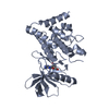 5hvjC  4bexS C: citing same article ( S: Starting model for refinement |
|---|---|
| Similar structure data |

-Links
 PDBj
PDBj


- Assembly
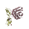
| Deposited unit | 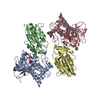
| ||||||||
|---|---|---|---|---|---|---|---|---|---|
| 1 | 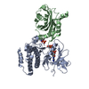
| ||||||||
| 2 | 
| ||||||||
| Unit cell |
|
-Components
| #1: Protein | Mass: 36473.191 Da / Num. of mol.: 2 / Fragment: UNP residues 329-638 / Mutation: D460N Source method: isolated from a genetically manipulated source Details: Optimized cDNA / Source: (gene. exp.) Homo sapiens (human) / Gene: LIMK1, LIMK / Production host:   Spodoptera frugiperda (fall armyworm) / Strain (production host): Sf9 Spodoptera frugiperda (fall armyworm) / Strain (production host): Sf9References: UniProt: P53667, non-specific serine/threonine protein kinase #2: Protein | | Mass: 18511.342 Da / Num. of mol.: 1 / Mutation: A69T Source method: isolated from a genetically manipulated source Source: (gene. exp.) Homo sapiens (human) / Gene: CFL1, CFL / Plasmid: pET-28a / Details (production host): N-terminal His6-SUMO tag / Production host:  #3: Protein | | Mass: 18431.363 Da / Num. of mol.: 1 / Mutation: A69T Source method: isolated from a genetically manipulated source Source: (gene. exp.) Homo sapiens (human) / Gene: CFL1, CFL / Plasmid: pET-28a / Details (production host): N-terminal His6-SUMO tag / Production host: #4: Chemical |   Mass: 506.196 Da / Num. of mol.: 2 / Source method: obtained synthetically / Formula: C10H17N6O12P3 / Comment: AMP-PNP, energy-carrying molecule analogue*YM Mass: 506.196 Da / Num. of mol.: 2 / Source method: obtained synthetically / Formula: C10H17N6O12P3 / Comment: AMP-PNP, energy-carrying molecule analogue*YM#5: Chemical |   Mass: 24.305 Da / Num. of mol.: 2 / Source method: obtained synthetically / Formula: Mg Mass: 24.305 Da / Num. of mol.: 2 / Source method: obtained synthetically / Formula: MgHas protein modification | Y | |
|---|
-Experimental details
-Experiment
| Experiment | Method: X-RAY DIFFRACTION / Number of used crystals: 1 |
|---|
- Sample preparation
| Crystal | Density Matthews: 2.68 Å3/Da / Density % sol: 54.08 % |
|---|---|
| Crystal grow | Temperature: 298 K / Method: vapor diffusion, hanging drop / pH: 5.5 Details: 1.2-1.4 M tri-Sodium citrate, 0.1 M sodium acetate, pH 5.5. 4 crystals used. |
-Data collection
| Diffraction | Mean temperature: 100 K |
|---|---|
| Diffraction source | Source: SYNCHROTRON / Site: APS / Beamline: 24-ID-E / Wavelength: 0.97918 Å |
| Detector | Type: ADSC QUANTUM 315 / Detector: CCD / Date: Mar 28, 2015 |
| Radiation | Protocol: SINGLE WAVELENGTH / Monochromatic (M) / Laue (L): M / Scattering type: x-ray |
| Radiation wavelength | Wavelength: 0.97918 Å / Relative weight: 1 |
| Reflection | Resolution: 3.5→50 Å / Num. obs: 15631 / % possible obs: 100 % / Redundancy: 10.1 % / Biso Wilson estimate: 33.7 Å2 / CC1/2: 0.531 / Rmerge(I) obs: 0.54 / Net I/σ(I): 2.5 |
| Reflection shell | Resolution: 3.5→3.63 Å / Redundancy: 8.5 % / Mean I/σ(I) obs: 1.3 / % possible all: 100 |
- Processing
| Software |
| |||||||||||||||||||||||||||||||||||||||||||||||||
|---|---|---|---|---|---|---|---|---|---|---|---|---|---|---|---|---|---|---|---|---|---|---|---|---|---|---|---|---|---|---|---|---|---|---|---|---|---|---|---|---|---|---|---|---|---|---|---|---|---|---|
| Refinement | Method to determine structure: MOLECULAR REPLACEMENT Starting model: 4BEX Resolution: 3.5→48.112 Å / SU ML: 0.52 / Cross valid method: FREE R-VALUE / σ(F): 1.33 / Phase error: 35.13 / Stereochemistry target values: ML
| |||||||||||||||||||||||||||||||||||||||||||||||||
| Solvent computation | Shrinkage radii: 0.9 Å / VDW probe radii: 1.11 Å / Solvent model: FLAT BULK SOLVENT MODEL | |||||||||||||||||||||||||||||||||||||||||||||||||
| Refinement step | Cycle: LAST / Resolution: 3.5→48.112 Å
| |||||||||||||||||||||||||||||||||||||||||||||||||
| Refine LS restraints |
| |||||||||||||||||||||||||||||||||||||||||||||||||
| LS refinement shell |
|
