[English] 日本語
Yorodumi
- PDB-5kzf: Crystal structure of near full-length hexameric Mycobacterium tub... -

+
Open data


ID or keywords:

Loading...

-
Basic information

Entry
Database: PDB / ID: 5kzf
Title
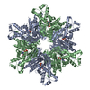
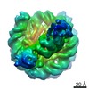
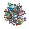
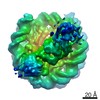
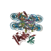
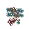
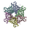
Crystal structure of near full-length hexameric Mycobacterium tuberculosis proteasomal ATPase Mpa in apo form
ComponentsProteasome-associated ATPase
KeywordsHYDROLASE / Proteasome
Function / homology
Function and homology information


proteasomal protein catabolic process / proteasome complex / modification-dependent protein catabolic process / ATP hydrolysis activity / ATP binding
Similarity search - Function
Proteasome ATPase / Proteasomal ATPase, N-terminal OB domain / Proteasomal ATPase OB N-terminal domain / Proteasomal ATPase OB C-terminal domain / Proteasomal ATPase OB C-terminal domain / ATPase, AAA-type, conserved site / AAA-protein family signature. / ATPase family associated with various cellular activities (AAA) / ATPase, AAA-type, core / ATPases associated with a variety of cellular activities ...Proteasome ATPase / Proteasomal ATPase, N-terminal OB domain / Proteasomal ATPase OB N-terminal domain / Proteasomal ATPase OB C-terminal domain / Proteasomal ATPase OB C-terminal domain / ATPase, AAA-type, conserved site / AAA-protein family signature. / ATPase family associated with various cellular activities (AAA) / ATPase, AAA-type, core / ATPases associated with a variety of cellular activities / AAA+ ATPase domain / P-loop containing nucleoside triphosphate hydrolase
Similarity search - Domain/homology
Proteasome-associated ATPase
Similarity search - Component
Biological speciesMycobacterium tuberculosis (bacteria)
MethodX-RAY DIFFRACTION / SYNCHROTRON / MOLECULAR REPLACEMENT / Resolution: 3.49 Å
AuthorsLi, H. / Hu, K. / Yang, S. / Bai, L.
Funding support United States, 1items
OrganizationGrant numberCountry
National Institutes of Health/National Institute Of Allergy and Infectious Diseases (NIH/NIAID)AI070285 United States
CitationJournal: Mol. Microbiol. / Year: 2017
Title: Mycobacterium tuberculosis proteasomal ATPase Mpa has a beta-grasp domain that hinders docking with the proteasome core protease.
Authors: Wu, Y. / Hu, K. / Li, D. / Bai, L. / Yang, S. / Jastrab, J.B. / Xiao, S. / Hu, Y. / Zhang, S. / Darwin, K.H. / Wang, T. / Li, H.
History
DepositionJul 25, 2016Deposition site: RCSB / Processing site: RCSB
Revision 1.0May 17, 2017Provider: repository / Type: Initial release
Revision 1.1Jul 12, 2017Group: Database references / Category: citation
Item: _citation.journal_volume / _citation.page_first / _citation.page_last
Revision 1.2Sep 20, 2017Group: Author supporting evidence / Category: pdbx_audit_support / Item: _pdbx_audit_support.funding_organization
Revision 1.3Dec 11, 2019Group: Advisory / Author supporting evidence / Derived calculations
Category: pdbx_audit_support / pdbx_validate_close_contact / struct_conn
Item: _pdbx_audit_support.funding_organization
Revision 1.4Mar 6, 2024Group: Data collection / Database references / Refinement description
Category: chem_comp_atom / chem_comp_bond ...chem_comp_atom / chem_comp_bond / database_2 / refine_hist
Item: _database_2.pdbx_DOI / _database_2.pdbx_database_accession / _refine_hist.d_res_low

-
Structure visualization

Structure viewerMolecule:
MolmilJmol/JSmol

Downloads & links

-
Assembly

Deposited unit
A: Proteasome-associated ATPase
B: Proteasome-associated ATPase
C: Proteasome-associated ATPase
D: Proteasome-associated ATPase
E: Proteasome-associated ATPase
F: Proteasome-associated ATPase
G: Proteasome-associated ATPase
H: Proteasome-associated ATPase
I: Proteasome-associated ATPase
J: Proteasome-associated ATPase
K: Proteasome-associated ATPase
L: Proteasome-associated ATPase


Theoretical massNumber of molelcules
Total (without water)682,15312
Polymers682,15312
Non-polymers00
Water00
1
A: Proteasome-associated ATPase
B: Proteasome-associated ATPase
C: Proteasome-associated ATPase
D: Proteasome-associated ATPase
E: Proteasome-associated ATPase
F: Proteasome-associated ATPase


Theoretical massNumber of molelcules
Total (without water)341,0776
Polymers341,0776
Non-polymers00
Water0
TypeNameSymmetry operationNumber
identity operation1_555x,y,z1
Buried area31560 Å2
ΔGint-79 kcal/mol
Surface area122500 Å2
MethodPISA
2
G: Proteasome-associated ATPase
H: Proteasome-associated ATPase
I: Proteasome-associated ATPase
J: Proteasome-associated ATPase
K: Proteasome-associated ATPase
L: Proteasome-associated ATPase


Theoretical massNumber of molelcules
Total (without water)341,0776
Polymers341,0776
Non-polymers00
Water0
TypeNameSymmetry operationNumber
identity operation1_555x,y,z1
Buried area31420 Å2
ΔGint-86 kcal/mol
Surface area124120 Å2
MethodPISA
Unit cell
Length a, b, c (Å)115.265, 202.590, 303.007
Angle α, β, γ (deg.)90.00, 90.00, 90.00
Int Tables number19
Space group name H-MP212121

-
Components

#1: Protein
Proteasome-associated ATPase / AAA ATPase forming ring-shaped complexes / ARC / Mycobacterial proteasome ATPase


Mass: 56846.094 Da / Num. of mol.: 12 / Fragment: residues 98-609
Source method: isolated from a genetically manipulated source
Source: (gene. exp.) Mycobacterium tuberculosis (strain ATCC 25177 / H37Ra) (bacteria)
Strain: ATCC 25177 / H37Ra / Gene: mpa, MRA_2130 / Production host: Escherichia coli (E. coli) / References: UniProt: A5U4E1

-
Experimental details

-
Experiment

ExperimentMethod: X-RAY DIFFRACTION / Number of used crystals: 1

-
Sample preparation

CrystalDensity Matthews: 2.6 Å3/Da / Density % sol: 52.68 %
Crystal growTemperature: 294 K / Method: vapor diffusion, hanging drop / pH: 7
Details: 100 mM Bis-Tris Propane, pH 7.0, 0.475 M NaCl, 18.2% PEG 4000, 20% glycerol

-
Data collection

DiffractionMean temperature: 100 K
Diffraction sourceSource: SYNCHROTRON / Site: NSLS / Beamline: X25 / Wavelength: 1.1 Å
DetectorType: ADSC QUANTUM 315 / Detector: CCD / Date: Aug 1, 2014
RadiationProtocol: SINGLE WAVELENGTH / Monochromatic (M) / Laue (L): M / Scattering type: x-ray
Radiation wavelengthWavelength: 1.1 Å / Relative weight: 1
ReflectionResolution: 3.49→71.129 Å / Num. obs: 90439 / % possible obs: 98.7 % / Redundancy: 3.1 % / Net I/σ(I): 9.9

-
Processing

Software
NameVersionClassification
REFMAC5.8.0123refinement
iMOSFLMdata reduction
SCALAdata scaling
PHASERphasing
RefinementMethod to determine structure: MOLECULAR REPLACEMENT / Resolution: 3.49→71.129 Å / Cor.coef. Fo:Fc: 0.903 / Cor.coef. Fo:Fc free: 0.872 / Cross valid method: THROUGHOUT / ESU R Free: 0.762 / Stereochemistry target values: MAXIMUM LIKELIHOOD / Details: HYDROGENS HAVE BEEN ADDED IN THE RIDING POSITIONS
RfactorNum. reflection% reflectionSelection details
Rfree0.30564 1996 2.2 %RANDOM
Rwork0.26772 ---
obs0.26856 88443 99.43 %-
Solvent computationIon probe radii: 0.8 Å / Shrinkage radii: 0.8 Å / VDW probe radii: 1.2 Å / Solvent model: MASK
Displacement parametersBiso mean: 128.92 Å2
Baniso -1Baniso -2Baniso -3
1--2.24 Å20 Å20 Å2
2--1.19 Å20 Å2
3---1.05 Å2
Refinement stepCycle: LAST / Resolution: 3.49→71.129 Å
ProteinNucleic acidLigandSolventTotal
Num. atoms44299 0 0 0 44299
Refine LS restraints
Refine-IDTypeDev idealDev ideal targetNumber
X-RAY DIFFRACTIONr_bond_refined_d0.0060.01945008
X-RAY DIFFRACTIONr_bond_other_d00.0243823
X-RAY DIFFRACTIONr_angle_refined_deg1.2011.98260865
X-RAY DIFFRACTIONr_angle_other_deg3.5553100983
X-RAY DIFFRACTIONr_dihedral_angle_1_deg6.36455633
X-RAY DIFFRACTIONr_dihedral_angle_2_deg36.89624.2262087
X-RAY DIFFRACTIONr_dihedral_angle_3_deg19.836158010
X-RAY DIFFRACTIONr_dihedral_angle_4_deg17.71515374
X-RAY DIFFRACTIONr_chiral_restr0.0530.26959
X-RAY DIFFRACTIONr_gen_planes_refined0.0050.02150509
X-RAY DIFFRACTIONr_gen_planes_other0.0020.029509
X-RAY DIFFRACTIONr_nbd_refined
X-RAY DIFFRACTIONr_nbd_other
X-RAY DIFFRACTIONr_nbtor_refined
X-RAY DIFFRACTIONr_nbtor_other
X-RAY DIFFRACTIONr_xyhbond_nbd_refined
X-RAY DIFFRACTIONr_xyhbond_nbd_other
X-RAY DIFFRACTIONr_metal_ion_refined
X-RAY DIFFRACTIONr_metal_ion_other
X-RAY DIFFRACTIONr_symmetry_vdw_refined
X-RAY DIFFRACTIONr_symmetry_vdw_other
X-RAY DIFFRACTIONr_symmetry_hbond_refined
X-RAY DIFFRACTIONr_symmetry_hbond_other
X-RAY DIFFRACTIONr_symmetry_metal_ion_refined
X-RAY DIFFRACTIONr_symmetry_metal_ion_other
X-RAY DIFFRACTIONr_mcbond_it4.1512.86322673
X-RAY DIFFRACTIONr_mcbond_other4.1512.86322672
X-RAY DIFFRACTIONr_mcangle_it6.21219.28428259
X-RAY DIFFRACTIONr_mcangle_other6.21219.28428260
X-RAY DIFFRACTIONr_scbond_it4.13913.0822335
X-RAY DIFFRACTIONr_scbond_other4.13913.0822336
X-RAY DIFFRACTIONr_scangle_it
X-RAY DIFFRACTIONr_scangle_other7.01519.53532607
X-RAY DIFFRACTIONr_long_range_B_refined9.23250755
X-RAY DIFFRACTIONr_long_range_B_other9.23150756
X-RAY DIFFRACTIONr_rigid_bond_restr
X-RAY DIFFRACTIONr_sphericity_free
X-RAY DIFFRACTIONr_sphericity_bonded
LS refinement shellResolution: 3.49→3.58 Å / Total num. of bins used: 20
RfactorNum. reflection% reflection
Rfree0.353 140 -
Rwork0.32 6198 -
obs--95.75 %

+
About Yorodumi

-
News

-
Feb 9, 2022. New format data for meta-information of EMDB entries

New format data for meta-information of EMDB entries

  • Version 3 of the EMDB header file is now the official format.
  • The previous official version 1.9 will be removed from the archive.

Related info.:EMDB header

External links:wwPDB to switch to version 3 of the EMDB data model

-
Aug 12, 2020. Covid-19 info

Covid-19 info

URL: https://pdbjlvh1.pdbj.org/emnavi/covid19.php

New page: Covid-19 featured information page in EM Navigator.

Related info.:Covid-19 info / Mar 5, 2020. Novel coronavirus structure data

+
Mar 5, 2020. Novel coronavirus structure data

Novel coronavirus structure data

Related info.:Yorodumi Speices / Aug 12, 2020. Covid-19 info

External links:COVID-19 featured content - PDBj / Molecule of the Month (242):Coronavirus Proteases

+
Jan 31, 2019. EMDB accession codes are about to change! (news from PDBe EMDB page)

EMDB accession codes are about to change! (news from PDBe EMDB page)

  • The allocation of 4 digits for EMDB accession codes will soon come to an end. Whilst these codes will remain in use, new EMDB accession codes will include an additional digit and will expand incrementally as the available range of codes is exhausted. The current 4-digit format prefixed with “EMD-” (i.e. EMD-XXXX) will advance to a 5-digit format (i.e. EMD-XXXXX), and so on. It is currently estimated that the 4-digit codes will be depleted around Spring 2019, at which point the 5-digit format will come into force.
  • The EM Navigator/Yorodumi systems omit the EMD- prefix.

Related info.:Q: What is EMD? / ID/Accession-code notation in Yorodumi/EM Navigator

External links:EMDB Accession Codes are Changing Soon! / Contact to PDBj

+
Jul 12, 2017. Major update of PDB

Major update of PDB

  • wwPDB released updated PDB data conforming to the new PDBx/mmCIF dictionary.
  • This is a major update changing the version number from 4 to 5, and with Remediation, in which all the entries are updated.
  • In this update, many items about electron microscopy experimental information are reorganized (e.g. em_software).
  • Now, EM Navigator and Yorodumi are based on the updated data.

External links:wwPDB Remediation / Enriched Model Files Conforming to OneDep Data Standards Now Available in the PDB FTP Archive

-
Yorodumi

Thousand views of thousand structures

  • Yorodumi is a browser for structure data from EMDB, PDB, SASBDB, etc.
  • This page is also the successor to EM Navigator detail page, and also detail information page/front-end page for Omokage search.
  • The word "yorodu" (or yorozu) is an old Japanese word meaning "ten thousand". "mi" (miru) is to see.

Related info.:EMDB / PDB / SASBDB / Comparison of 3 databanks / Yorodumi Search / Aug 31, 2016. New EM Navigator & Yorodumi / Yorodumi Papers / Jmol/JSmol / Function and homology information / Changes in new EM Navigator and Yorodumi

Read more