 Movie
Movie Controller

[English] 日本語
 Yorodumi
- PDB-3hyv: 3-D X-Ray structure of the sulfide:quinone oxidoreductase from th... -
+ Open data

- Basic information
| Entry | Database: PDB / ID: 3hyv | |||||||||
|---|---|---|---|---|---|---|---|---|---|---|
| Title | 3-D X-Ray structure of the sulfide:quinone oxidoreductase from the hyperthermophilic bacterium Aquifex aeolicus | |||||||||
 Components Components | Sulfide-quinone reductase | |||||||||
 Keywords Keywords | OXIDOREDUCTASE / PROTEIN COMPLEX / MONOTOPIC MEMBRANE PROTEIN / FLAVOPROTEIN / ROSSMANN-FOLD DOMAIN | |||||||||
| Function / homology |  Function and homology information Function and homology informationbacterial sulfide:quinone reductase / sulfide:quinone oxidoreductase activity / NAD(P)H dehydrogenase (quinone) activity / aerobic electron transport chain / quinone binding / nucleotide binding / membrane / identical protein binding Similarity search - Function | |||||||||
| Biological species |   Aquifex aeolicus (bacteria) Aquifex aeolicus (bacteria) | |||||||||
| Method |  X-RAY DIFFRACTION / SYNCHROTRON / MIRAS / Resolution: 2.3 Å X-RAY DIFFRACTION / SYNCHROTRON / MIRAS / Resolution: 2.3 Å | |||||||||
 Authors Authors | Marcia, M. / Ermler, U. / Peng, G.H. / Michel, H. | |||||||||
 Citation Citation | Journal: Proc.Natl.Acad.Sci.USA / Year: 2009 Title: The structure of Aquifex aeolicus sulfide:quinone oxidoreductase, a basis to understand sulfide detoxification and respiration Authors: Marcia, M. / Ermler, U. / Peng, G.H. / Michel, H. | |||||||||
| History |
|
- Structure visualization
| Structure viewer | Molecule: MolmilJmol/JSmol |
|---|
- Downloads & links
-Download
| PDBx/mmCIF format | 3hyv.cif.gz | 521.6 KB | Display | PDBx/mmCIF format |
|---|---|---|---|---|
| PDB format | pdb3hyv.ent.gz | 429.8 KB | Display | PDB format |
| PDBx/mmJSON format | 3hyv.json.gz | Tree view | PDBx/mmJSON format | |
| Others |  Other downloads Other downloads |
-Validation report
| Arichive directory | https://data.pdbj.org/pub/pdb/validation_reports/hy/3hyvftp://data.pdbj.org/pub/pdb/validation_reports/hy/3hyv | HTTPS FTP |
|---|
-Related structure data


-Links
 PDBj
PDBj


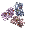
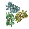
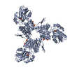
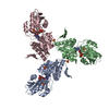
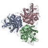
- Assembly
| Deposited unit | 
| ||||||||
|---|---|---|---|---|---|---|---|---|---|
| 1 | 
| ||||||||
| 2 | 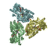
| ||||||||
| 3 | 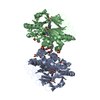
| ||||||||
| 4 | 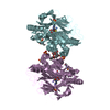
| ||||||||
| 5 | 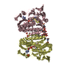
| ||||||||
| Unit cell |
|
-Components
-Protein / Sugars , 2 types, 12 molecules ABCDEF
| #1: Protein | Mass: 47534.918 Da / Num. of mol.: 6 / Source method: isolated from a natural source / Source: (natural) Aquifex aeolicus (bacteria) / Strain: VF5References: UniProt: O67931, Oxidoreductases; Acting on a sulfur group of donors; With a quinone or similar compound as acceptor #3: Sugar | ChemComp-LMT /  Type: D-saccharide / Mass: 510.615 Da / Num. of mol.: 6 / Source method: obtained synthetically / Formula: C24H46O11 / Comment: detergent*YM Type: D-saccharide / Mass: 510.615 Da / Num. of mol.: 6 / Source method: obtained synthetically / Formula: C24H46O11 / Comment: detergent*YM |
|---|
-Non-polymers , 6 types, 620 molecules 
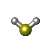


| #2: Chemical | ChemComp-FAD /  Mass: 785.550 Da / Num. of mol.: 6 / Source method: obtained synthetically / Formula: C27H33N9O15P2 / Comment: FAD*YM Mass: 785.550 Da / Num. of mol.: 6 / Source method: obtained synthetically / Formula: C27H33N9O15P2 / Comment: FAD*YM#4: Chemical | ChemComp-H2S /  Mass: 34.081 Da / Num. of mol.: 6 / Source method: obtained synthetically / Formula: H2S Mass: 34.081 Da / Num. of mol.: 6 / Source method: obtained synthetically / Formula: H2S#5: Chemical | ChemComp-PS9 /  Mass: 256.520 Da / Num. of mol.: 7 / Source method: obtained synthetically / Formula: S8 Mass: 256.520 Da / Num. of mol.: 7 / Source method: obtained synthetically / Formula: S8#6: Chemical | ChemComp-MES /  Mass: 195.237 Da / Num. of mol.: 6 / Source method: obtained synthetically / Formula: C6H13NO4S / Comment: pH buffer*YM Mass: 195.237 Da / Num. of mol.: 6 / Source method: obtained synthetically / Formula: C6H13NO4S / Comment: pH buffer*YM#7: Chemical | ChemComp-SO4 /  Mass: 96.063 Da / Num. of mol.: 24 / Source method: obtained synthetically / Formula: SO4 Mass: 96.063 Da / Num. of mol.: 24 / Source method: obtained synthetically / Formula: SO4#8: Water | ChemComp-HOH / | Mass: 18.015 Da / Num. of mol.: 571 / Source method: isolated from a natural source / Formula: H2O |
|---|
-Details
| Has protein modification | Y |
|---|---|
| Nonpolymer details | THE SIDE CHAIN OF CYS156 IS EXTENDED TO FORM A PUTATIVE POLYSULFUR CHAIN. ONE SULFUR ATOM OF THE ...THE SIDE CHAIN OF CYS156 IS EXTENDED TO FORM A PUTATIVE POLYSULFUR |
-Experimental details
-Experiment
| Experiment | Method: X-RAY DIFFRACTION / Number of used crystals: 1 |
|---|
- Sample preparation
| Crystal | Density Matthews: 2.73 Å3/Da / Density % sol: 54.92 % |
|---|---|
| Crystal grow | Temperature: 291 K / Method: vapor diffusion, hanging drop / pH: 6.5 Details: 4% PEG 400 (V/V), 0.1M NA-MES (SODIUM 2-(N-MORPHOLINO)-ETHANESULFONATE), 2M AMMONIUM SULFATE, pH 6.5, VAPOR DIFFUSION, HANGING DROP, temperature 291K |
-Data collection
| Diffraction |
| ||||||||||||||||||
|---|---|---|---|---|---|---|---|---|---|---|---|---|---|---|---|---|---|---|---|
| Diffraction source |
| ||||||||||||||||||
| Detector |
| ||||||||||||||||||
| Radiation |
| ||||||||||||||||||
| Radiation wavelength |
| ||||||||||||||||||
| Reflection | Resolution: 2.3→50 Å / Num. obs: 135807 / % possible obs: 97.9 % / Redundancy: 3.47 % / Net I/σ(I): 14.87 | ||||||||||||||||||
| Reflection shell | Resolution: 2.3→2.4 Å / Redundancy: 2.47 % / Mean I/σ(I) obs: 3.44 / % possible all: 87.9 |

- Processing
| Software |
| ||||||||||||||||||||||||||||||||||||||||||||||||||||||||||||||||||||||||||||||||||||||||||||||||||||||||||||||||||||||||||||||||||||||||||||||||||||||||||||||||||||||||||
|---|---|---|---|---|---|---|---|---|---|---|---|---|---|---|---|---|---|---|---|---|---|---|---|---|---|---|---|---|---|---|---|---|---|---|---|---|---|---|---|---|---|---|---|---|---|---|---|---|---|---|---|---|---|---|---|---|---|---|---|---|---|---|---|---|---|---|---|---|---|---|---|---|---|---|---|---|---|---|---|---|---|---|---|---|---|---|---|---|---|---|---|---|---|---|---|---|---|---|---|---|---|---|---|---|---|---|---|---|---|---|---|---|---|---|---|---|---|---|---|---|---|---|---|---|---|---|---|---|---|---|---|---|---|---|---|---|---|---|---|---|---|---|---|---|---|---|---|---|---|---|---|---|---|---|---|---|---|---|---|---|---|---|---|---|---|---|---|---|---|---|---|
| Refinement | Method to determine structure: MIRAS / Resolution: 2.3→20.04 Å / Cor.coef. Fo:Fc: 0.95 / Cor.coef. Fo:Fc free: 0.926 / SU B: 13.13 / SU ML: 0.165 / Cross valid method: THROUGHOUT / ESU R: 0.322 / ESU R Free: 0.226 / Stereochemistry target values: MAXIMUM LIKELIHOOD / Details: HYDROGENS HAVE BEEN ADDED IN THE RIDING POSITIONS
| ||||||||||||||||||||||||||||||||||||||||||||||||||||||||||||||||||||||||||||||||||||||||||||||||||||||||||||||||||||||||||||||||||||||||||||||||||||||||||||||||||||||||||
| Solvent computation | Ion probe radii: 0.8 Å / Shrinkage radii: 0.8 Å / VDW probe radii: 1.2 Å / Solvent model: MASK | ||||||||||||||||||||||||||||||||||||||||||||||||||||||||||||||||||||||||||||||||||||||||||||||||||||||||||||||||||||||||||||||||||||||||||||||||||||||||||||||||||||||||||
| Displacement parameters | Biso mean: 38.517 Å2
| ||||||||||||||||||||||||||||||||||||||||||||||||||||||||||||||||||||||||||||||||||||||||||||||||||||||||||||||||||||||||||||||||||||||||||||||||||||||||||||||||||||||||||
| Refinement step | Cycle: LAST / Resolution: 2.3→20.04 Å
| ||||||||||||||||||||||||||||||||||||||||||||||||||||||||||||||||||||||||||||||||||||||||||||||||||||||||||||||||||||||||||||||||||||||||||||||||||||||||||||||||||||||||||
| Refine LS restraints |
| ||||||||||||||||||||||||||||||||||||||||||||||||||||||||||||||||||||||||||||||||||||||||||||||||||||||||||||||||||||||||||||||||||||||||||||||||||||||||||||||||||||||||||
| LS refinement shell | Resolution: 2.3→2.359 Å / Total num. of bins used: 20
|
