 Movie
Movie Controller

[English] 日本語
 Yorodumi
- PDB-5ksr: Stationary phase survival protein E (SurE) from Xylella fastidios... -
+ Open data

- Basic information
| Entry | Database: PDB / ID: 5ksr | ||||||
|---|---|---|---|---|---|---|---|
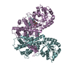
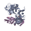
| Title | Stationary phase survival protein E (SurE) from Xylella fastidiosa - XFSurE-TB (Tetramer Bigger). | ||||||
 Components Components | 5'-nucleotidase SurE | ||||||
 Keywords Keywords | HYDROLASE / Stationary phase survival protein E (SurE) / Xylella fastidiosa / crystallization. | ||||||
| Function / homology |  Function and homology information Function and homology information3'-nucleotidase activity / exopolyphosphatase activity / 5'-nucleotidase / 5'-nucleotidase activity / nucleotide binding / metal ion binding / identical protein binding / cytoplasm Similarity search - Function | ||||||
| Biological species |  Xylella fastidiosa (bacteria) Xylella fastidiosa (bacteria) | ||||||
| Method |  X-RAY DIFFRACTION / SYNCHROTRON / MOLECULAR REPLACEMENT / Resolution: 1.96 Å X-RAY DIFFRACTION / SYNCHROTRON / MOLECULAR REPLACEMENT / Resolution: 1.96 Å | ||||||
 Authors Authors | Machado, A.T.P. / Fonseca, E.M.B. / Dos Reis, M.A. / Saraiva, A.M. / Dos Santos, C.A. / De Toledo, M.A. / Polikarpov, I. / De Souza, A.P. / De Aparicio, R. / Iulek, J. | ||||||
| Funding support |  Brazil, 1items Brazil, 1items
| ||||||
 Citation Citation | Journal: Proteins / Year: 2017 Title: Conformational variability of the stationary phase survival protein E from Xylella fastidiosa revealed by X-ray crystallography, small-angle X-ray scattering studies, and normal mode analysis. Authors: Machado, A.T.P. / Fonseca, E.M.B. / Reis, M.A.D. / Saraiva, A.M. / Santos, C.A.D. / de Toledo, M.A.S. / Polikarpov, I. / de Souza, A.P. / Aparicio, R. / Iulek, J. | ||||||
| History |
|
- Structure visualization
| Structure viewer | Molecule: MolmilJmol/JSmol |
|---|
- Downloads & links
-Download
| PDBx/mmCIF format | 5ksr.cif.gz | 415.5 KB | Display | PDBx/mmCIF format |
|---|---|---|---|---|
| PDB format | pdb5ksr.ent.gz | 340.6 KB | Display | PDB format |
| PDBx/mmJSON format | 5ksr.json.gz | Tree view | PDBx/mmJSON format | |
| Others |  Other downloads Other downloads |
-Validation report
| Arichive directory | https://data.pdbj.org/pub/pdb/validation_reports/ks/5ksrftp://data.pdbj.org/pub/pdb/validation_reports/ks/5ksr | HTTPS FTP |
|---|
-Related structure data
| Related structure data |  5ksqC 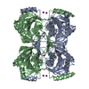 5kssC  5kstC 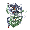 3ty2S C: citing same article ( S: Starting model for refinement |
|---|---|
| Similar structure data |


-Links
 PDBj
PDBj- Assembly
| Deposited unit | 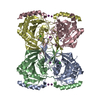
| ||||||||
|---|---|---|---|---|---|---|---|---|---|
| 1 |
| ||||||||
| Unit cell |
| ||||||||
| Components on special symmetry positions |
|
-Components
| #1: Protein | Mass: 29490.037 Da / Num. of mol.: 4 Source method: isolated from a genetically manipulated source Source: (gene. exp.) Xylella fastidiosa (strain 9a5c) (bacteria)Strain: 9a5c / Gene: surE, XF_0858 / Production host: #2: Chemical | ChemComp-MN /   Mass: 54.938 Da / Num. of mol.: 4 / Source method: obtained synthetically / Formula: Mn Mass: 54.938 Da / Num. of mol.: 4 / Source method: obtained synthetically / Formula: Mn#3: Chemical | ChemComp-IOD /   Mass: 126.904 Da / Num. of mol.: 6 / Source method: obtained synthetically / Formula: I Mass: 126.904 Da / Num. of mol.: 6 / Source method: obtained synthetically / Formula: I#4: Chemical | ChemComp-CL /   Mass: 35.453 Da / Num. of mol.: 4 / Source method: obtained synthetically / Formula: Cl Mass: 35.453 Da / Num. of mol.: 4 / Source method: obtained synthetically / Formula: Cl#5: Water | ChemComp-HOH / |  Mass: 18.015 Da / Num. of mol.: 661 / Source method: isolated from a natural source / Formula: H2O Mass: 18.015 Da / Num. of mol.: 661 / Source method: isolated from a natural source / Formula: H2O |
|---|
-Experimental details
-Experiment
| Experiment | Method: X-RAY DIFFRACTION / Number of used crystals: 1 |
|---|
- Sample preparation
| Crystal | Density Matthews: 2.77 Å3/Da / Density % sol: 55.61 % |
|---|---|
| Crystal grow | Temperature: 293 K / Method: vapor diffusion, hanging drop Details: 0.1 M bis-tris propane pH 7.5, 0.14 M Sodium iodide, 20% (W/V) PEG 3350, 5 mM dithiotheitol and 0.1 manganese II chloride. |
-Data collection
| Diffraction | Mean temperature: 100 K |
|---|---|
| Diffraction source | Source: SYNCHROTRON / Site: LNLS / Beamline: W01B-MX2 / Wavelength: 1.459 Å |
| Detector | Type: MARMOSAIC 225 mm CCD / Detector: CCD / Date: Jul 15, 2008 |
| Radiation | Protocol: SINGLE WAVELENGTH / Monochromatic (M) / Laue (L): M / Scattering type: x-ray |
| Radiation wavelength | Wavelength: 1.459 Å / Relative weight: 1 |
| Reflection | Resolution: 1.96→43.248 Å / Num. all: 88536 / Num. obs: 88536 / % possible obs: 99.5 % / Observed criterion σ(I): -3 / Redundancy: 4.77 % / Biso Wilson estimate: 29.72 Å2 / Rmerge(I) obs: 0.049 / Net I/σ(I): 17.27 |
| Reflection shell | Resolution: 1.96→2.02 Å / Redundancy: 4.34 % / Rmerge(I) obs: 0.338 / Mean I/σ(I) obs: 4.8 / % possible all: 95.7 |
- Processing
| Software |
| |||||||||||||||||||||||||||||||||||||||||||||||||||||||||||||||||||||||||||||||||||||||||||||||||||||||||||||||||||||||||||||||||||||||||||||||||||||||||||||||||||||||||||||||||||||||||||||||||||||||||||||||||||||||||
|---|---|---|---|---|---|---|---|---|---|---|---|---|---|---|---|---|---|---|---|---|---|---|---|---|---|---|---|---|---|---|---|---|---|---|---|---|---|---|---|---|---|---|---|---|---|---|---|---|---|---|---|---|---|---|---|---|---|---|---|---|---|---|---|---|---|---|---|---|---|---|---|---|---|---|---|---|---|---|---|---|---|---|---|---|---|---|---|---|---|---|---|---|---|---|---|---|---|---|---|---|---|---|---|---|---|---|---|---|---|---|---|---|---|---|---|---|---|---|---|---|---|---|---|---|---|---|---|---|---|---|---|---|---|---|---|---|---|---|---|---|---|---|---|---|---|---|---|---|---|---|---|---|---|---|---|---|---|---|---|---|---|---|---|---|---|---|---|---|---|---|---|---|---|---|---|---|---|---|---|---|---|---|---|---|---|---|---|---|---|---|---|---|---|---|---|---|---|---|---|---|---|---|---|---|---|---|---|---|---|---|---|---|---|---|---|---|---|---|
| Refinement | Method to determine structure: MOLECULAR REPLACEMENT Starting model: 3TY2 Resolution: 1.96→43.248 Å / SU ML: 0.22 / Cross valid method: FREE R-VALUE / Phase error: 25.07
| |||||||||||||||||||||||||||||||||||||||||||||||||||||||||||||||||||||||||||||||||||||||||||||||||||||||||||||||||||||||||||||||||||||||||||||||||||||||||||||||||||||||||||||||||||||||||||||||||||||||||||||||||||||||||
| Solvent computation | Shrinkage radii: 1.1 Å / VDW probe radii: 1.4 Å | |||||||||||||||||||||||||||||||||||||||||||||||||||||||||||||||||||||||||||||||||||||||||||||||||||||||||||||||||||||||||||||||||||||||||||||||||||||||||||||||||||||||||||||||||||||||||||||||||||||||||||||||||||||||||
| Displacement parameters | Biso mean: 36.04 Å2 | |||||||||||||||||||||||||||||||||||||||||||||||||||||||||||||||||||||||||||||||||||||||||||||||||||||||||||||||||||||||||||||||||||||||||||||||||||||||||||||||||||||||||||||||||||||||||||||||||||||||||||||||||||||||||
| Refinement step | Cycle: LAST / Resolution: 1.96→43.248 Å
| |||||||||||||||||||||||||||||||||||||||||||||||||||||||||||||||||||||||||||||||||||||||||||||||||||||||||||||||||||||||||||||||||||||||||||||||||||||||||||||||||||||||||||||||||||||||||||||||||||||||||||||||||||||||||
| Refine LS restraints |
| |||||||||||||||||||||||||||||||||||||||||||||||||||||||||||||||||||||||||||||||||||||||||||||||||||||||||||||||||||||||||||||||||||||||||||||||||||||||||||||||||||||||||||||||||||||||||||||||||||||||||||||||||||||||||
| LS refinement shell |
| |||||||||||||||||||||||||||||||||||||||||||||||||||||||||||||||||||||||||||||||||||||||||||||||||||||||||||||||||||||||||||||||||||||||||||||||||||||||||||||||||||||||||||||||||||||||||||||||||||||||||||||||||||||||||
| Refinement TLS params. | Method: refined / Refine-ID: X-RAY DIFFRACTION
| |||||||||||||||||||||||||||||||||||||||||||||||||||||||||||||||||||||||||||||||||||||||||||||||||||||||||||||||||||||||||||||||||||||||||||||||||||||||||||||||||||||||||||||||||||||||||||||||||||||||||||||||||||||||||
| Refinement TLS group |
|
